MIC Symposium 2022
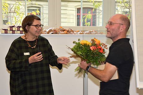
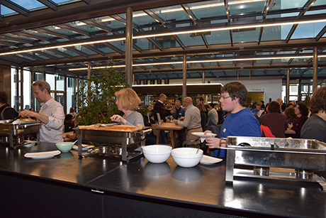
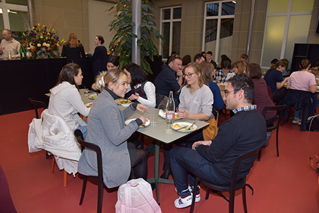
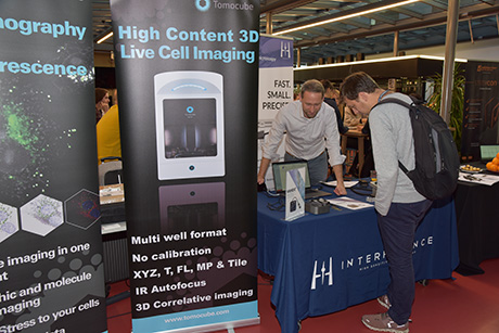
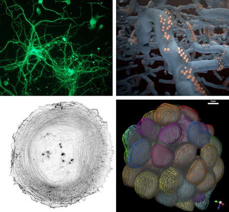
On November 18, 2022, the MIC Symposium “Imaging Cellular Dynamics Across Scales” celebrated how new microscopy technologies redefine our understanding of dynamic cellular processes at different biological scales. The different talks illustrated how each biological scale requires a complementary set of microscope technology, specific image analysis algorithms, as well as a variety of genetically encoded fluorescent probes (GFP derivatives). Francesca Peri kicked off the meeting by showing beautiful cell biology data of how macrophages constantly remodel the microglia in the central nervous system by very dynamic interactions. Thanks to light sheet microscopy this can now be studied directly in the zebrafish brain. Suliana Manley then showed how new super resolution microscopy techniques can provide new insight in the cellular machineries that allow for mitochondria fission and fusion. Of particular interest, the “event driven acquisition” intelligent microscopy modality can now automatically recognize cellular events of interest, to subsequently study them at high temporal resolution without a user input. Miki Ebisuya then explained how a stem cell zoo allows her team to explore the regulation of the segmentation clock that explains the different gestation times of the zoo’s species. She also showed how to manipulate the mechanical processes contributing to development using optogenetics in organoids. Joachim Goedhart then explained how the large number of fluorescent proteins, designed to tackle different imaging tasks, were rationally engineered. He also explained which considerations one must think about selecting the best fluorescent protein to image one’s favorite biological process. Olivier Pertz then discussed how the MAPK signaling pathway operates in space and time in single living cells. He showed how simple cell communication rules of signaling allow a cell ecosystem to coordinate proliferation, survival, apoptosis and migration fates. He then explained how these cell communication rules are coopted in cancer to coordinate aberrant invasion and proliferation. Christian Soeller showed how the ryanodine receptor is organized at the nanometer scale to regulate the calcium flux underlying many physiological processes. Further, company talks displayed the latest microscopy technologies aimed at visualizing different biological scales. Before the lunch break, two PhD students that graduated from the PhD program “Cutting Edge Microscopy” received their certificate and Olivier Pertz was warmly thanked for his many years of service on the MIC board. The symposium ran very well due to excellent preparation by the MIC administration and the scientific committee and great support by our student helpers who made sure that all speakers’ and audience needs were catered for. During the breaks, people gathered in the foyer for lively discussions over food and drink. The industrial stands invited visitors to find out about industry developments. In the end, it was a spectacular day with stunning movies that documented how biological processes can be extremely entertaining!
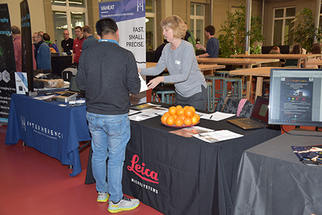
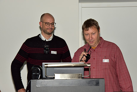
Impressions
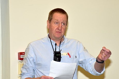
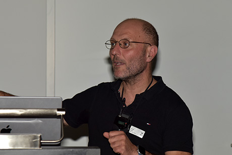
Speaker
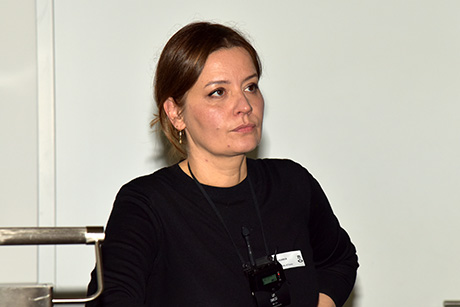
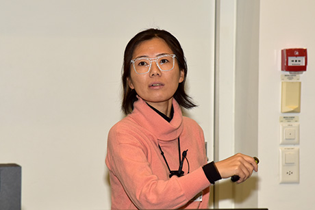
PhD Miki Ebisuya, European Molecular Biology Laboratory (EMBL), Barcelona, Spain
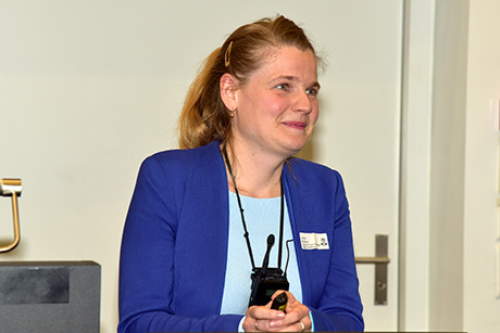
Prof. Dr. Suliana Manley, Department of Physics, Swiss Federal Institute of Technology Lausanne (EPFL), Switzerland
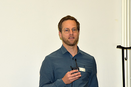
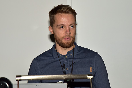
Dr. ir Joachim Goedhart, Swammerdam Institute for Life Sciences, The Netherlands
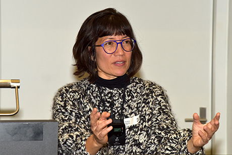
Prof. Dr. Francesca Peri, Department of Molecular Life Sciences, University of Zurich, Switzerland
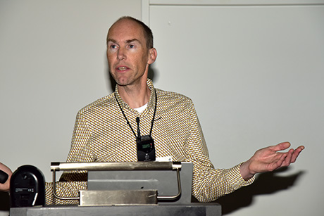
Prof. Dr. Olivier Pertz, Institute of Cell Biology, University of Bern, Switzerland
PhD Arina Rybina, Euro-BioImaging Bio-Hub, European Molecular Biology Laboratory (EMBL), Heidelberg, Germany
Prof. Dr. Christian Soeller, Institute of Physiology, University of Bern, Switzerland
Supported by
- Abberior
- Agilent Technologies
- GMP General Microtechnology
- Hamamatsu
- Interherence
- Leica
- Nikon
- Prospective Instruments
- SVI
- Tomocube
- Witec
- Zeiss
Impressions