News
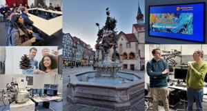
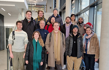
Study Trip 2026, CEM PhD Program, Göttingen, Germany
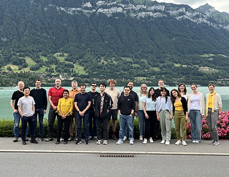
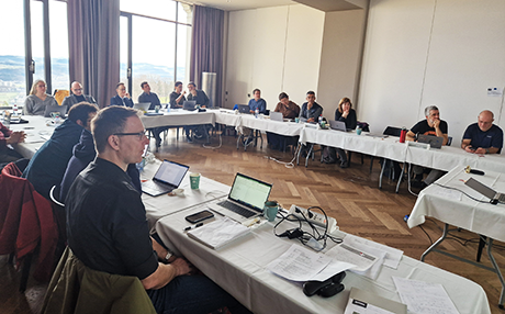
From January 21–23, 2026, the PhD students of the Cutting Edge Microscopy specialization program from the University of Bern visited Göttingen, Germany, for an intensive scientific program focused on state-of-the-art fluorescence microscopy, instrument development, and imaging workflows in biomedical research. The visit combined industrial perspectives, academic research, and facility-level discussions, offering a comprehensive view of advanced microscopy technologies.
The program began with a visit to abberior Instruments GmbH, where participants received an in-depth introduction to STED-based super-resolution microscopy. Technical presentations and live demonstrations highlighted key aspects of optical design, system stability, and practical trade-offs between resolution and imaging speed. The day concluded with a public lecture by Nobel Laureate Stefan Hell at the Max Planck Institute, who placed super-resolution microscopy into a broader conceptual and historical context and discussed future challenges in nanoscopy, including labelling strategies and biological interpretation at nanometer scales.
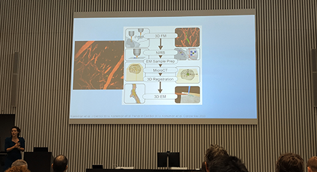
The second day focused on the development and application of advanced microscopy workflows in biomedical research. At the University Medical Centre Göttingen, Fred Wouters and Gertrude Bunt presented imaging approaches aimed at modernising pathology workflows, including rapid tissue clearing for light-sheet microscopy and the integration of correlated light and electron microscopy (CLEM) for the analysis of neurodegenerative disease samples. In the afternoon, the group visited the Georg-August University Göttingen, where Jan Huisken introduced cutting-edge light-sheet microscopy technologies. A live demonstration of the Flamingo light-sheet system illustrated how interdisciplinary collaboration enables long-term, high-speed imaging of living specimens with minimal photodamage.
The final day took place at the Max Planck Institute within Peter Lénárt’s group and focused on microscopy as a centralised research service. Discussions covered a wide range of imaging modalities as well as organisational and strategic aspects of operating a modern microscopy facility. The visit concluded with the return journey to Bern, providing time for reflection on the scientific and institutional insights gained during the three days.
Read the students' report below:
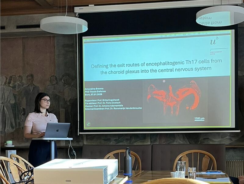
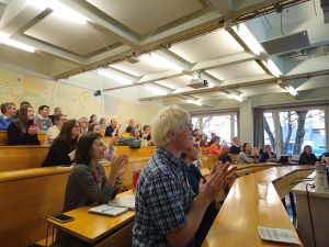
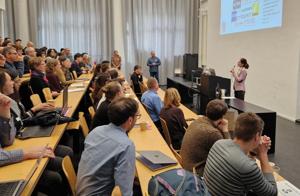
Congratulations on the successful PhD Defence of Amandine Brenna
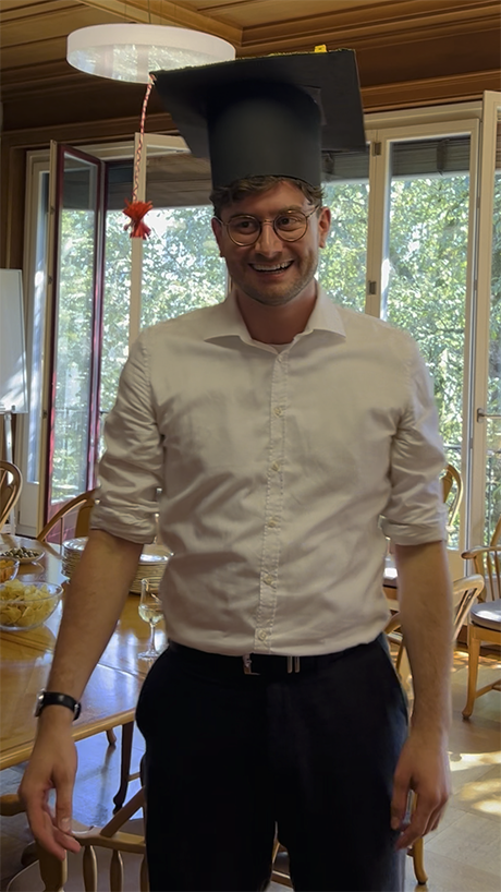
On January 7, 2026, Amandine Brenna, student of the Cutting Edge Microscopy (CEM) program under the supervision of Prof. Dr. Britta Engelhardt (UniBE) and Prof. Dr. Fiona Doetsch (UniBasel), successfully defended her PhD thesis with the title “Defending the exist routes of encephalitogenic Th17 cells from the choroid plexus into the central nervous system.”
Amandine reports this from her thesis: “During my PhD, I investigated how encephalitogenic Th17 cells access the central nervous system (CNS) via the choroid plexus stroma during experimental autoimmune encephalomyelitis (EAE). To define the anatomical exit routes used by Th17 cells, we first examined the organization of the choroid plexus and surrounding meningeal structures. We then developed a fluorescent reporter mouse model enabling visualization of the blood-cerebrospinal fluid barrier of the choroid plexus, in which encephalitogenic Th17 cells are transferred to induce EAE. By combining two-photon intravital microscopy, confocal imaging, and whole-brain light-sheet fluorescence microscopy, our findings suggest that Th17 cells may use an as-yet-undiscovered route to invade the CNS during neuroinflammation.”
Electron Microscopy MIC Mini Symposium 2025
Tuesday, 2025/12/09, 15:00
The MIC Mini Symposium marked the official inauguration of the new scanning and transmission electron microscopes at the Vetsuisse Faculty. The event featured a series of engaging presentations demonstrating the wide range of applications of electron microscopy, including animal hair in an archaeological context, bacteriophages diversity, apicomplexan parasites, the use of EM in fish and crayfish health, and the visualization of bacteria.
We would like to extend our deepest gratitude to everyone who made this event possible — from the dedicated MIC administration team and Veterinary Anatomy team to the funding bodies, as well as all speakers and attendees whose enthusiasm and support contributed to a lively and inspiring atmosphere.
The symposium was very well received, with stimulating talks highlighting both the scientific diversity and the impact of electron microscopy. Overall, the event was a success and provided a welcome platform for scientific exchange, inspiration, and future collaborations.
We hope all attendees enjoyed the program and gained insight into electron microscopy applications that enable a more detailed look into cells, microorganisms, viruses, nanoparticles, and surface structures.
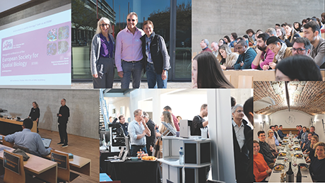
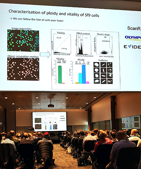
MIC Symposium 2025: Exploring "Multi-Scale Imaging Across Modalities"
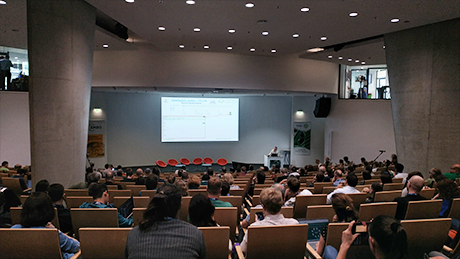
The annual Microscopy Imaging Center (MIC) Symposium, held on November 14, 2025, celebrated how cutting-edge technologies are advancing our ability to visualize biological structures and function across a vast range of resolutions.
This year's theme, “Multi-Scale Imaging Across Modalities,” showcased state-of-the-art technical advancements and emerging techniques essential for modern biomedical research. The day was structured into three main sessions, covering the entire imaging scale from high-resolution electron microscopy to novel optical approaches and X-ray microtomography.
1. Possibilities of 3-Dimensional Electron Microscopy
The day kicked off with a Welcome from Nadia Mercader, UniBe’s Medical Facultys Vice Dean. The first main session was chaired by Ruslan Hlushchuk. Benoît Zuber (University of Bern) started the session by outlining practical strategies, tips, and tricks for optimizing Serial Block-Face Scanning Electron Microscopy (SBF-SEM). Next, Christel Genoud (University of Lausanne and EPFL) provided an overview of volume electron microscopy (volume EM) techniques on their platform, showing how they bridge the gap between ultrastructural detail and large-volume context. Kedar Narayan (National Cancer Institute, NIH, USA) concluded the session, focusing on moving "Towards truly quantitative volume electron microscopy" by leveraging AI-based solutions for large-scale segmentation of cellular ultrastructure.
2. Advancements in Optical Microscopic Techniques
Chaired by Britta Engelhardt, this session explored the latest in optical imaging. Laura Batti (Wyss Center for Neuroimaging and Neurobiology, Geneva) highlighted how Light-Sheet Microscopy is empowering advancements in neuroscience and medicine, allowing for imaging of entire, cleared organs with high resolution. Following this, Christian Soeller (University of Bern) presented the MIC's recently established super-resolution pipeline, illustrating its practical applications from the micrometer to the single nanometer scale. Isto Kristian Jänönen (Thermo Fisher Scientific) gave a presentation on the Invitrogen EVOS S1000 Spatial Imaging System and its role in multiplexed IHC workflows for spatial biology.
3. Emerging Techniques for Biological Imaging
The final session, chaired by Steven Proulx, focused on novel imaging modalities. Ivano Romano (Evident Europe) detailed new developments in confocal microscopy, including improvements in speed, sensitivity, and the game-changing dynamic range of the Fluoview4000 system. Robert Prevedel (EMBL Heidelberg, Germany) discussed "Unconventional optical approaches," including advancements in Brillouin microscopy for capturing cellular and tissue visco-elasticity. The symposium concluded with Georg Schulz (University of Basel), who illuminated the potential of cellular-resolution X-ray microtomography for non-destructive biomedical research, achieving voxel sizes down to 180nm.
Industry Exhibition and Organization
Before the lunch break, a certificate award was presented to the graduates of the CEM PhD program: Jana Leuenberger, Oleksiy Khoma and Mohammadamin Khosrozadeh. Throughout the day, attendees engaged in lively discussions over food and drink, both during the lunch break and dedicated coffee breaks. The industrial exhibition allowed visitors to find out about the latest industry developments, thanks to the generous support of sponsors, including Zeiss, Evidents, Nikon, Thermo Fisher Scientific, and Vitaris.
The MIC Symposium was once again a great success, running smoothly thanks to the MIC administration team and the scientific committee: Britta Engelhardt, Ruslan Hlushchuk, and Steven Proulx. The event was also approved for a 0.5-day credit for continued education in animal experimentation in the Canton of Bern.
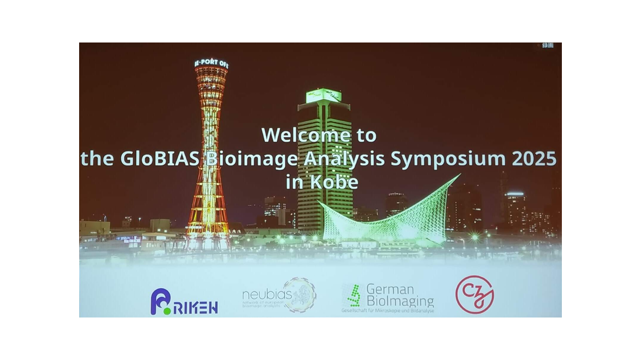
GloBIAS Bioimage Analysis Symposium 2025 in Kobe, Japan
The GloBIAS Bioimage Analysis Symposium 2025 took place from 29–31 October 2025 at RIKEN BDR in Kobe, Japan. The symposium was part of a larger event that also included a training school, hackathons, and a taggathon. It brought together around 150 researchers, software developers, and imaging specialists from around the world to advance open and reproducible bioimage analysis.
The scientific programme featured invited talks, poster presentations, and interactive sessions such as the Open-Source Software Lounge and Call-for-Help forum. Topics ranged from open-source tool development and FAIR data principles to machine learning, workflow management, and quantitative imaging. The symposium emphasised collaboration between life scientists, computer scientists, and data engineers in order to strengthen the global bioimage analysis community.
The Microscopy Imaging Center (MIC) was represented by Yury Belyaev, who presented the poster Benchmarking deconvolution tools for microscopy core facilities, co-authored with Diego Morone (Institute for Research in Biomedicine, Bellinzona) and Felix Meyenhofer (University of Fribourg). Yury also visited colleagues from imaging facilities at the National Institute for Basic Biology in Okazaki and the RIKEN Center for Biosystems Dynamics Research in Kobe.

Congratulations to two successful CEM PhD Defenses
In the last two months, two PhD students from the Cutting Edge Microscopy (CEM) program successfully defended their theses. Congratulations!
Oleksiy Khoma
On August 22, 2025, Oleksiy Khoma, student of the Cutting Edge Microscopy (CEM) program under the supervision of PD Dr. Ruslan Hlushchuk and Dr. David Haberthür, successfully defended his PhD thesis with the title “Micro(angio)CT in preclinical dental research.”
Oleksiy reports this from his thesis: “During my PhD at the University of Bern, I developed and validated a microangioCT-based framework for three-dimensional, non-destructive visualization of peri-implant vascular networks in large animal models. Using the polymer-based contrast agent μAngiofil, I optimized perfusion techniques and imaging protocols to capture microvascular architecture around dental implants with high spatial resolution. The methodology bridges the gap between traditional histology and advanced 3D imaging, enabling quantitative analysis of peri-implant soft tissue vascularization and advancing preclinical research toward improved dental implant integration and tissue regeneration strategies.”
Jana Leuenberger
On September 30, 2025, Jana Leuenberger, student of the Cutting Edge Microscopy (CEM) program under the supervision of Prof. Dr. Benoît Zuber, successfully defended her PhD thesis entitled “Integrative Analysis of Synaptic Structure and Function in PC12, SH-SY5Y, and iPSC-derived Motor Neurons: A Multimodal Evaluation of Model Characteristics.”
Jana reports this from her thesis: During my PhD I explored how different neuronal cell models establish and maintain synaptic structures and functions. Using a combination of fluorescence microscopy, live-cell imaging, electrophysiology, conventional and cryo-electron microscopy, I characterized synapse formation, vesicle recycling, and ultrastructural organization across several neuronal systems. My work revealed distinct stages of synaptic maturation, from the assembly of presynaptic and postsynaptic protein complexes to the emergence of functional vesicle recycling and activity-dependent plasticity. By integrating fluorescence imaging with cryo-electron tomography, I visualized synaptic vesicle pools, connectors, and active-zone architectures at nanometer resolution, providing an integrated view of how molecular organization relates to synaptic function in human-derived neurons. These results contribute to a deeper mechanistic understanding of synapse formation and offer a framework for selecting and validating cellular models for high-resolution structural and functional human studies of neuronal connectivity.

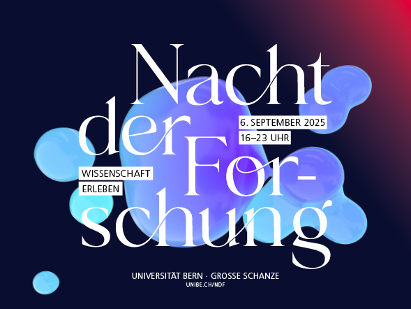
MIC at the «Nacht der Forschung» 2025
On Saturday, 6th September, the University of Bern hosted the "Nacht der Forschung" (Night of Research), a university-wide event that transformed the campus into a "festival of knowledge". The Microscopy Imaging Center (MIC) was one of over 80 projects that welcomed approximately 10,000 visitors, inviting them on a journey into the microcosm. Over 50 dedicated volunteers, a mix of students and staff, guided guests through the secrets of animal and plant cells.
The MIC’s exhibit provided an artistic and hands-on look at research in the life sciences. Interactive stations allowed guests to use professional microscopes to view hidden life and fascinating structures. Attendees could also learn how to prepare their own samples and even fold an origami microscope to take home. For a deeper dive, visitors experienced a new perspective through virtual reality, traveling through the delicate skeleton of a zebrafish or the internal structures of a mouse's skull. The event also featured a "Sci-Art" gallery where aesthetic images merged with biological knowledge. Thanks to a loan from the Institute for the History of Medicine, the MIC was able to showcase an exhibition of historical microscopes.
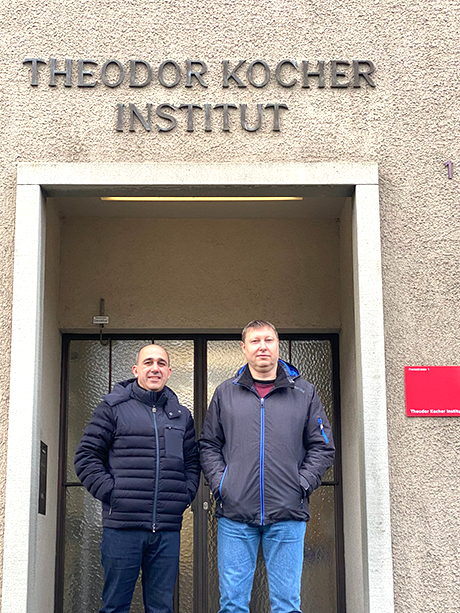
The night showcased the collaborative spirit of the university, featuring projects from various institutes, including the Institute of Plant Sciences, the Theodor Kocher Institute, and the Institute of Anatomy. The enthusiastic and curious faces of children and adults alike were a testament to the night's success. The MIC team extends a heartfelt thank you to all the volunteers who made their part of the event possible.
Eine Reise in den Mikrokosmos
Die Geheimnisse pflanzlicher und tierischer Zellen
Wie Pflanzen atmen, bleibt unserem Auge meist verborgen – winzige Spaltöffnungen, feine Zellstrukturen und lebendige Oberflächen entziehen sich dem bloßen Blick. Doch unter dem Mikroskop wird das Unsichtbare sichtbar. Das Microscopy Imaging Center (MIC) macht genau diese Wunderwelt für alle zugänglich: Mit Mikroskopie-Stationen, eigenen Präparaten, Origami Mikroskopen und Virtual-Reality-Rundgängen lädt es Neugierige, Lernende und Fachpersonen jeden Alters ein, die Welt des Kleinen zu entdecken – interaktiv, anschaulich und inspirierend.
Wir werden im 2. Stock des Hauptgebäudes der Universität Bern sein, Hochschulstrasse 4, 3012 Bern - Wir freuen uns auf Besuchende!
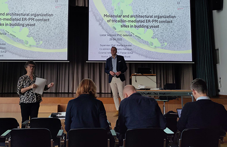
Congratulations to successful PhD Defence of Lazar Ivanovic
On April 29, 2025, Lazar Ivanovnic, student of the Cutting Edge Microscopy (CEM) program under the supervision of Prof. Wanda Kukulski, successfully defended his PhD thesis with the title “Molecular and architectural organization of tricalbin-mediated ER-PM contact sites in budding yeast.”
What Lazar reports from his thesis: During my PhD I investigated the molecular and architectural organization of the lipid transfer proteins tricalbins at endoplasmic reticulum (ER) and plasma membrane (PM) contact sites in yeast cells. By employing fluorescence recovery after photobleaching (FRAP) we uncover that tricalbin heteromerization renders ER membrane curvature sensitivity which slows down their diffusional mobility and promotes accumulation at ER-PM contact sites. Furthermore, we piece information on temporal dynamics together with ultrastructural insights from correlative light and electron microscopy (CLEM). Our results show that intermembrane distance as well as large intrinsically disordered regions control the cellular distribution and dynamic behaviour of tricalbins in cells.
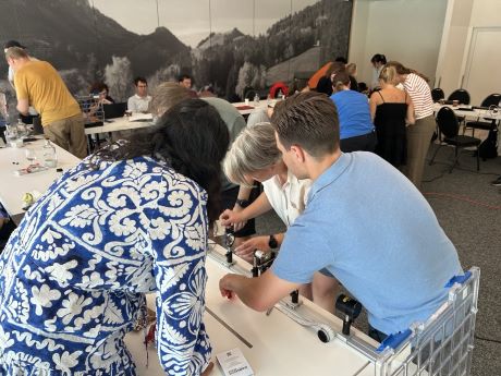
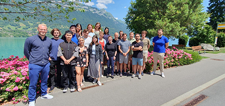
CEM Summer School 2025
On the 3rd and 4th of July 2025, the annual Summer School of the PhD specialization program Cutting Edge Microscopy (CEM) took place at Schwarzsee in the canton of Fribourg, Switzerland. During these two days the CEM PhD students presented their own projects, focusing on the data obtained with microscopy. In lively discussions they received feedback and gained fresh perspectives on their research.
The students’ presentations were accompanied by talks from senior researchers Jens Stein (University of Fribourg, UniFR) and Maria Rosito (Link Campus University, Italy), who shared valuable insights from their projects. Ana Stojiljković and Ruben Lopez (Data Science Lab, UniBE) presented the progress of the VIBE platform (UniBE) and how it will support image analysis in the future.
There was interactive and creative fun with the “Build Your Own Microscope” challenge, led by Head of Bioimage core facility Boris Egger and Bioimage Analyst Felix Meyenhofer (both UniFR). To clear heads and enjoy the beautiful alpine scenery the students (and professors) got to go on a 4km Monster-Trottinett ride down the Riggisalp — an unforgettable experience for all.
Read more about the two Summer School days in the PDF below, written by the students.
MIC Research Day 2025
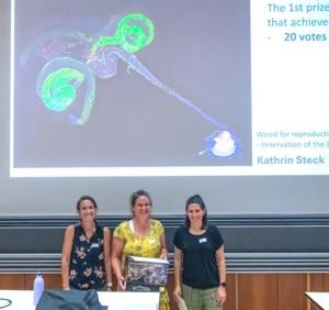
On June 25, 2025, more than 180 participants from the Universities of Bern, Fribourg, Zürich and Bellinzona as well as representatives from the industry attended our traditional MIC Research Day. The balanced program of the event was presented by microscopists Thomas Auer, Soheila Zeinali, Amandine Brenna, Andrew Hemphill, Leonie Anton, Gaelle Lentini, Aurella Emonet, Marina Eckermann and Alexandre Bokhobza, ranging from PhD students to established researchers and coming from all three faculties of the MIC (Vetsuisse, Natural Sciences and Medicine) as well as our guest Maria Rosito from the Link Campus University in Rome. In excellent presentations, these scientists showed the broad spectrum of microscopy applications represented in the MIC community.
A highlight of this year’s event was the newly introduced Image contest, where participants were asked to send in their most beautiful, funny and thought-provoking pictures. After a communal vote the 3 winners got their well-deserved prizes! Thanks to a generous contribution from 2 of our sponsors, the first prize was a Nikon Camera, the second a Huygens-Pro subscription from Huygens. In addition, Guillaume Witz and his team introduced the VIBE Platform, a Bioimage Analysis Platform which will serve the microscopy community of the University of Bern by fall 2026. Participants were invited to the MIC Symposium 2025 on November 14 in Bern, with this year’s topic "Multi-Scale Imaging Across Modalities".
As usual, this event offered numerous opportunities for networking among microscopists at the University of Bern. Networking was also supported by the industry partners with their exhibition and industry talk, which fitted well into the event. We would like to thank all CEM PhD students for their help with the organization, all participants for their enthusiasm for microscopy and last but not least the event sponsors for their generous financial support.
Have a look at some impressions from the MIC Research Day 2025 here.
International European Light Microscopy Initiative Meeting (ELMI) 2025
The 25th International European Light Microscopy Initiative Meeting (ELMI) took place from June 3–6, 2025, at EMBL Heidelberg, Germany, marking the initiative’s 25th anniversary. The event opened with the traditional Core Facility Day on June 3. Over the next two and a half days, the scientific programme featured lectures by leading scientists and invited speakers from across Europe and beyond which showcased the latest research and technological developments in light microscopy.
Session topics included probes and biosensors, smart and advanced microscopy, super-resolution imaging, tissue imaging and light sheet microscopy, imaging for health, and data analysis. In parallel, hands-on workshops and industry-led training offered practical insights into cutting-edge technologies and applications.
The conference brought together around 600 participants, with approximately 500 from academia and 100 from industry. More than 50 companies in microscopy and related fields presented their latest innovations through technical workshops and exhibitions.
The Microscopy Imaging Center (MIC) was represented by Yury Belyaev, who presented the poster Boosting precision in cryo-CLEM via deconvolution, developed together with Jaime Llodrá and Benoît Zuber (also a MIC member) from the Institute of Anatomy, University of Bern.
Yury Belyaev at Stellenbosch University, South Africa
In April 2025, Yury Belyaev, Light Microscopy Core Facility Manager at the Microscopy Imaging Center (MIC) of the University of Bern, visited the Central Analytical Facilities (CAF) Microscopy Unit at Stellenbosch University, South Africa (https://www.sun.ac.za/english/research-innovation/caf/units-laboratories/microscopy). The visit was part of the Global BioImaging (GBI) Job Shadowing Program (https://globalbioimaging.org/international-job-shadowing), which fosters international knowledge exchange and skills development among imaging core facilities worldwide.
The primary objective of this visit was to gain deeper insights into the organization, workflows, and technical expertise of the CAF Microscopy Unit, while also sharing the best practices in core facility management and microscopy services. Throughout the program, Yury Belyaev observed and discussed various aspects of imaging facility operations, including user training, sample preparation, service provision, data management, and advanced imaging techniques.
Yury was warmly welcomed by the CAF team and received a comprehensive tour of the microscopy platform. Particular attention was given to light and electron microscopy as well as correlative imaging approaches. The exchange involved fruitful discussions on workflow optimization, strategies for user support, and the integration of new technologies into routine services.
During his stay, Yury Belyaev also took part in a microscopy workshop organized by the CAF Microscopy Unit. This hands-on training session brought together participants from across South Africa and focused on wide-field and confocal microscopy, image analysis, and microscope maintenance. The workshop was additionally supported by the CoRE Genomics for Health in Africa Scientific Exchange Program (https://www.genomicsforhealthinafrica.org/sciex-registration), which provided travel grants for three participants, thereby enhancing accessibility and regional collaboration.
Yury expresses his sincere gratitude to the entire CAF Microscopy team, especially Unit Manager Lize Engelbrecht, for their hospitality and willingness to share their expertise. He also extends thanks to Global BioImaging for enabling this enriching professional exchange and for their generous financial support.
Congratulations to PhD graduation of CEM-program member Mohammad Amin Khosrozadeh
On January 30, 2025, Mohammad Amin Khosrozadeh, member of the Institute of Anatomy, Research Group of Prof. Dr. Benoît Zuber, University of Bern, and student at the Graduate School for Cellular and Biomedical Sciences (GCB) and the PhD program Cutting Edge Microscopy (CEM), has successfully defended his PhD thesis entitled "Morphological Principle of Synaptic Transmission Regulation Using Cryo-Electron Tomography and Data-Driven Models".
Dr. Khosrozadeh reports on his research as follows: During my PhD studies, I investigated the nanoscale organization of synaptic vesicles and their regulation using advanced cryo-electron tomography (cryo-ET) techniques and computational analysis methods. I developed CryoVesNet, a deep learning-based segmentation tool that enables automated analysis of synaptic vesicles in their native state. By applying graph theoretical modeling to synaptic ultrastructure, I uncovered how vesicle pools transition between supercritical and critical states during varying levels of synaptic activity. This work provides new insights into the structural basis of neurotransmission and synaptic plasticity. Additionally, I established methodologies for high-resolution structural analysis of membrane proteins, achieving sub-4Å resolution of pore-forming toxins while preserving their native lipid interactions. These approaches open new possibilities for understanding the molecular architecture of synapses and the mechanisms underlying neurological disorders.
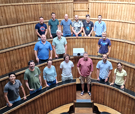
PhD program Cutting Edge Microscopy - Study Trip 2025
On January 24 and 25, 2025, a study tour took 10 PhD students of the PhD specialization program Cutting Edge Microscopy (CEM) and senior researchers involved in steering the CEM program to Heidelberg, Germany. On the first day, the students visited the Infectious Diseases Imaging Platform (IDIP) at the Center for Integrative Infectious Disease (CIID) at Heidelberg University and on the second day, the study tour continued with a visit to the brand new Imaging Center at the European Molecular Biology Laboratory (EMBL). Both facilities have an impressive portfolio of imaging techniques and microscopic instruments. Educational presentations with research examples from institute members and site visits provided an insight into the facilities. The institute directors Vibor Laketa, IDIP, Heidelberg University, and Timo Zimmermann, Imaging Center, EMBL, warmly welcomed the students and guided them through the program. The event was rounded off with an interesting guided tour of Heidelberg at night. This news article is supplemented by a downloadable report written by the students.
MIC Symposium 2024
On November 15th, the MIC Symposium themed “Bio-inspired materials and bioengineering” was hosted for the first time at the University of Fribourg. Science Faculty Vice Dean, Barbara Rothen-Rutishauser, welcomed about 150 participants in the Joseph Deiss Auditorium. She highlighted microscopy at the University of Fribourg and as the first MIC coordinator shared insights about the early days of the MIC in Bern.
Simon Sprecher from Biology, University of Fribourg, kicked off the biomedical imaging session, demonstrating how neuronal activity can be visualized in Drosophila taste organs. Anja Hauser from Charité Berlin discussed how the tissue environment influences the metabolism of myeloid cells in bone marrow. Christian Soeller from Physiology, University of Bern, presented super-resolved images of receptor clusters in cardiac muscle cells using the new MINFLUX system. Closing the session, Mario Prsa from Medicine, University of Fribourg, showed a robotic system that delivers precise proprioceptive stimuli to a mouse forelimb while imaging and manipulating neuronal brain activity with multiphoton microscopy.
During lunch and coffee breaks, attendees interacted with 14 commercial exhibitors showcasing the latest technologies and instruments.
The second session focused on how natural phenomena inspire the development of innovative materials and technologies. Aleksandra Radenovic from EPFL used fluorescence microscopy to monitor electrochemical reactions at the single-molecule scale. Derek Kiebala from Chemistry, University of Mainz, demonstrated the use of confocal microscopy to study force sensors at the microscale level. Viola Bauernfeind from AMI, University of Fribourg, concluded the session by showing how the order or disorder of microscopic structures affects structural colors in nature.
The third session addressed advances in imaging single molecules at nanoscale. Guillermo Acuna from Phyisics, University of Fribourg, presented a self-made low-cost super-resolution microscope adapted for smartphones. Pablo Rivera-Fuentes from the University of Zürich showed how the blinking patterns of single fluorophores can be used to accurately distinguish peptide sequences. Sophie Brasselet from the Institute Fresnel in Marseille concluded the symposium by demonstrating how the 3D orientation of single molecules can be detected in cells using polarization fluorescence microscopy.
The local scientific committee, Barbara Rothen-Rutishauser, Jens Stein, Boris Egger, and Dimitri Vanhecke, extends their gratitude to all speakers for their insightful and inspiring talks. Special thanks go to the MIC administration and local crew for their meticulous preparations. The symposium would not be possible without the generous financial support from the numerous commercial sponsors, thank you all! Finally, thanks to all participants for the lively interactive sessions. We hope you enjoyed the MIC symposium and look forward to seeing you again next year!

SNSF Scientific Image Competition 2025
Get your cameras! Give Swiss research a face.
The SNSF Scientific Image Competition encourages researchers working in Switzerland to present their works to the public and the media. Photographs, images and videos will be rated in terms of their aesthetic quality and their ability to inspire and amaze, to convey or illustrate knowledge, to tell a human story or to let us discover a new universe.
The winning images will be exhibited at the Biel/Bienne Festival of Photography in May 2025.
Submission deadline is January 31, 2025.
Find more information here.
Congratulations to successful PhD Defense Adrian Madarasz
On July 9, 2024, Adrian Madarasz, student of the Cutting Edge Microscopy (CEM) program under the supervision of Dr. Steven Proulx, has successfully defended his PhD thesis with the title: "Pathways and Dynamcis of Erythrocyte Efflux in Murine Models of Hemorrhagic Stroke".
During my PhD studies, I explored the involvement of the lymphatic system regarding the outflow of red blood cells (RBCs) in murine hemorrhagic stroke models. Employing newly developed labeling procedures for RBCs combined with epifluorescence in vivo imaging and 3D reconstructions from confocal microscopy of decalcified tissue, we have shown that the lymphatic vessels, especially in the cribriform plate region, contribute significantly to the clearance of RBCs from the subarachnoid space. Contrary to previous publications, access of RBCs to dorsal dural lymphatics was limited at early time points. In addition, we compared different models for intracerebral hemorrhage and their suitability for studying the efflux of cerebrospinal fluid or tracers from the subarachnoid space.
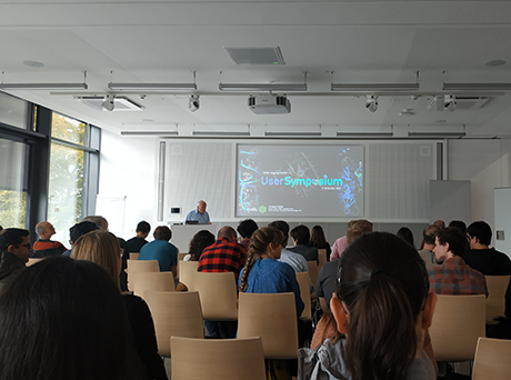
EMBL Imaging Center (EIC) Imaging Symposium
The EMBL Imaging Centre (EIC) User Symposium (https://www.embl.org/about/info/imaging-centre/32247-2/) was held on October 17-18, 2024, at EMBL Heidelberg, Germany. This event brought together around 60 current and prospective users of EIC (https://www.embl.org/about/info/imaging-centre/), along with leading experts in electron and light microscopy.
The symposium was preceded by workshops from Leica and Zeiss, where both companies showcased their latest products and services. Following the workshops, participants engaged in an extensive two-day scientific program. Sessions on light and electron microscopy featured presentations from field experts and EIC users, who shared insights from their research. Additionally, a short-talk session provided attendees with information about the services offered by the EIC and its future plans.
The symposium also served as an excellent networking platform for microscopy enthusiasts from various research institutes in Europe. During a panel discussion moderated by Simone Mattei and Timo Zimmermann (team leaders of electron and light microscopy at the EIC, respectively), participants had the opportunity to exchange experiences using the EIC’s services. Yury Belyaev from MIC also attended the meeting.
Researchers from the University of Bern can access the services of the EMBL Imaging Centre, as Switzerland is a member state of EMBL. Those interested in taking advantage of this opportunity should reach out via email at ic-contact@embl.de.
European Microscopy Congress (EMC) 2024
The 17th European Microscopy Congress (EMC) took place on August 24-29, 2024, in Copenhagen, Denmark, attracting over 1,500 researchers and industry representatives. The event focused on the latest advancements in microscopy in the life and materials sciences, with a strong emphasis on both light and electron microscopy techniques.
The EMC 2024 included sessions on light and electron microscopy, correlative methods, novel imaging modalities, and image processing. The event’s highlights were its plenary talks, five parallels symposia, and industry-led workshops. Attendees also explored emerging microscopy technologies, participated in interactive sessions, and attended pre-conference workshops.
In addition to the scientific program, over 90 leading manufacturers and suppliers of microscopy instrumentation showcased their latest products. This interaction between academia and industry offered delegates valuable insights into cutting-edge tools. The exhibition space fostered collaboration opportunities and supported the adoption of innovative technologies within the research community.
The Microscopy Imaging Center (MIC) was represented by Benoit Zuber and Yury Belyaev at EMC 2024. Calvin Klein, a PhD student from Wanda Kukulski’s lab and a member of the CEM program, also attended and received the best poster presentation award.
Open positions MIC & DSL
There are currently two open positions in MIC and DSL. Please spread the word if you know someone who might be interested and don't hesitate to contact us or apply if you are interested in either position. You can find the vacancies here: Bioimage Analyst for new platform / IT Specialist for Bioimage Analysis Platform
The Microscopy Imaging Center (MIC) and the Data Science Lab (DSL) are core facilities of the University of Bern providing scientific support for researchers across all faculties. The two units have long-standing collaboration to help researchers access advanced image analysis methods through custom software development and support for existing bioimage analysis tools.
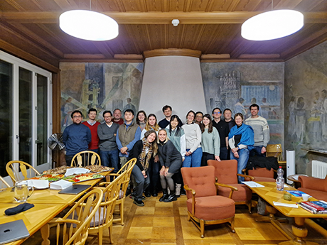
CEM Summer School 2024
From July 4 to 5, 2024, the annual summer school of the PhD program Cutting Edge Microscopy (CEM) took place at the Seehotel in Bönigen, Switzerland. All students gave scientific presentations on their projects, focusing on applied microscopy and the results obtained. To enhance the interactive style of the presentations, the students presented in teams. The teams had to find a common denominator, usually the research area, which they then presented in the form of a joint presentation. This format encouraged a more lively atmosphere and was much appreciated by all participants. The subsequent individual progress reports from the students delved deeper into the microscopy applications. The review of the scientific part of the summer school took the form of a quiz where each student contributed to a pool of questions. Senior participants of the CEM Summer School 2024 were MIC microscopy specialists Fabian Blank and Yury Belyaev, MIC bioimaging specialist Ana Stojiljkovic from the Data Science Lab (DSL) and CEM PhD program scientific co-directors Benoît Zuber and Steven Proulx. Ruth Lyck, the coordinator of the CEM PhD program, was responsible for the quiz and the organization of the trip. A special guest was Vibor Laketa, Head of the Platform for Infectious Disease Imaging at the University of Heidelberg, who gave a lecture on the topic of ethics in handling and publishing BioImage data. In addition to the scientific program, a discussion about the upcoming activities of the CEM PhD program took place and the new CEM student representatives Marwa Mangattu Parambil and Jasmina El Fata were elected. The beautiful landscape, the pleasant accommodation and the free time to enjoy Lake Brienz or to go jogging contributed to a perfect learning atmosphere.
MIC Research Day 2024
On June 26, 2024, more than 180 participants from the Universities of Bern, Fribourg and Zurich, the EPFL in Lausanne and representatives from industry attended the traditional MIC Research Day, which took place at the Department of Chemistry, Biochemistry and Pharmaceutical Sciences (DCBP). The balanced program of the event was presented by microscopists Li Xin, Isabel Schultz-Pernice, Saiko Yoshida, Oleksiy-Zakhar Khoma, Alicia Borgeaud, Divyansh Gautam, Matis Preza, Reto Lang and Benjamin Towbin, ranging from PhD students to established researchers and coming from all three faculties of the MIC (Vetsuisse, Natural Sciences and Medicine). In excellent presentations, these scientists showed the broad spectrum of microscopy applications represented in the MIC community.
In addition, Peter Gehr, Professor Emeritus of the Institute of Anatomy and co-founder of the MIC, spoke about the history of the MIC. Two students of the PhD specialization program Cutting Edge Microscopy (CEM) presented the key elements of the CEM program. Dimitri Vanhecke invited the MIC community to the MIC Symposium 2024 on November 15 in Fribourg, with the theme "Bio-inspired Materials & Bio-Engineering". Arne Seitz from the Microscopy Facility of the EPFL promoted the LS2 intersection Microscopy, which forms an organ of the Swiss microscopy community. Martin Frenz and Volker Heussler were officially thanked for their many years of service as MIC committee members, Volker Heussler also being a member of the MIC board for several years. Our special guest Nicholas James Desnoyer from the University of Zurich gave a breathtaking presentation on the development of plants in live imaging. Nicholas wowed the MIC community with his enthusiasm and dedication to showing the beauty of nature in pictures and movies.
As usual, this event offered numerous opportunities for networking among imaging professionals at the University of Bern. To encourage networking, a meet-the-expert session was offered during lunch. Networking was also supported by the industry partners with their exhibition and industry talk, which fitted well into the event. We would like to thank all CEM PhD students for their help with the organization, all participants for their enthusiasm for microscopy and the event sponsors for their generous financial support.
Please find some impressions from the MIC Research Day 2024 here.
Congratulations to successful PhD Defense Javier Pareja Román
On June 6, 2024 Javier Pareja Román, student of the Cutting Edge Microscopy (CEM) program under the supervision of Prof. Britta Engelhardt, has successfully defended his PhD thesis with the title: "Unveiling the cellular and molecular pathways of CD8 T cell migration into the CNS"
During my PhD I have been investigating different antigen dependent and antigen independent mechanisms that regulate the migration of CD8 T cells across the different CNS barriers during health and neuroinflammation. In particular we have shown that CD8 T cells arrested on the blood-brain barrier (BBB) can recognised antigens presented on the luminal surface of the endothelium. These interactions halt the CD8 T cells and reduce their transmigration, eventually leading to BBB breakdown contributing to the exacerbation of neuroinflammation.
In addition, I have intensely used different microscopy techniques to characterise novel reporter mouse models for intravital imaging of the meninges at the surface of the CNS and the glia limitans, with the goal of investigating in vivo the trafficking of CD8 T cells across this CNS barriers.
European Light Microscopy Initiative Meeting (ELMI) 2024
The 23rd International European Light Microscopy Initiative Meeting (ELMI) took place from June 4-7, 2024, in Liverpool, UK, gathering over 600 researchers, scientists and industry representatives to discuss the latest advancements in light microscopy, focusing on life sciences.
The event started with a Core Facility Day, featuring sessions on tools and resources, data management and spatial omics. Core facility members from different countries shared their experiences. For instance, Nicholas Condon from the University of Queensland in Australia discussed the implementation of the Image Processing Platform and Laurent Gelman from FMI in Switzerland highlighted various tools and resources used in microscopy facilities. A community discussion concluded the session, summarized the tools and resources presented.
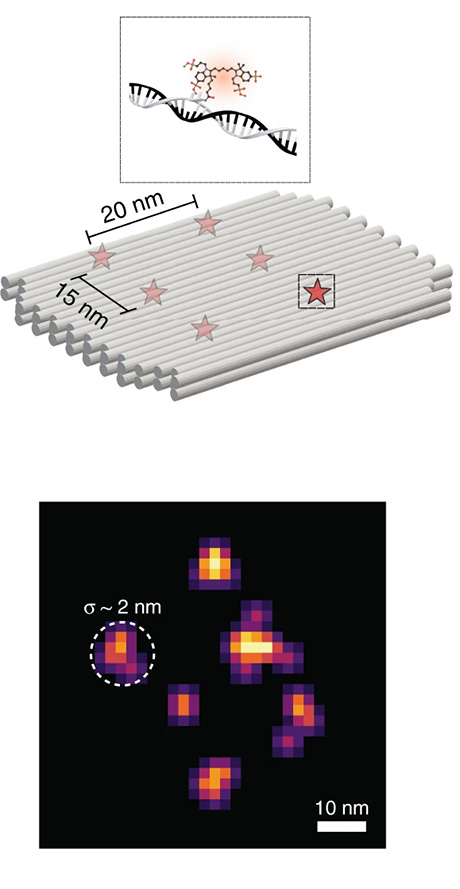
Over the next two and a half days, leading scientists presented their latest research and innovations in microscopy. Topics included multimodal correlative imaging, image analysis, intelligent acquisition, super-resolution microscopy and imaging across scales. These presentations emphasized significant advancements and future directions in microscopy, highlighting the field's dynamic and rapidly evolving nature. Notably, Ralf Jungmann from the Max Planck Institute of Biochemistry delivered a keynote lecture on spatial omics using DNA-based super-resolution microscopy.
In addition to scientific presentations, more than 50 leading companies in microscopy instrumentation and related fields showcased their latest products and technologies through informative workshops and booth presentations. This interaction between academia and industry provided attendees with insights into cutting-edge tools and techniques, fostering potential collaborations and technological adoption.
The Microscopy Imaging Center (MIC) was represented by Yury Belyaev. Overall, ELMI 2024 provided an excellent platform for networking, knowledge exchange and fostering collaborations between industry and academia.
Zeiss User Meeting 2024
In collaboration with the Microscopy Imaging Center the Zeiss User Meeting 2024 will take place at the University of Bern.
Selected Highlights:
- Swiss Premiere of the newest ZEISS Microscope: Lattice SIM 3.
- Inspiring Talks (SMLM, Structured Illumination, Multiplex Imaging, and much more)
- Networking in the Swiss Life Science Community during Apero and Dinner
When: 3 September 2024 - Start at 10.00 a.m., open end with dinner at 19.00 p.m.
Where: Institute of Cell Biology, Baltzerstrasse 4, University of Bern
Register soon as places are limited, register here.
Find more information here.
Industry call for the best microscopy image
Following the success of past Image of the Year Awards, the search for the best light microscopy images in 2024 continues. The fifth annual Image of the Year competition recognizes the very best in scientific imaging worldwide.
The contest welcomes materials science images in addition to life science images to show the versatility of the art of science. This year, a new video category is introduced, enabling microscopists to show off the best of their moving images.
Participants can win an SZX7 microscope with a DP23 digital camera, X Line™ objectives, a CX23 microscope, or an SZ61 microscope.
Click here to learn more and submit your entry. Good luck!
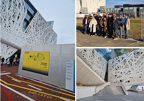
CEM Study Trip 2024
On February 1 and 2, 2024, a study trip took 18 PhD students of the PhD specialization program Cutting Edge Microscopy (CEM) and 3 "seniors", who were the CEM program scientific co-director Benoît Zuber, the MIC light microscopy specialist Yury Belyaev and the CEM coordinator Ruth Lyck, to Milan, Italy. There, the students visited the Human Technopole Institute (HT) in Rho Fiera. The HT is a large-scale research infrastructure launched by the Italian government in 2018. Since 2024, the HT has opened the first national facilities offering services and training for biomedical research with cutting-edge technology. Fabrizio Martino, Licensing Officer at the Human Technopole, warmly welcomed the students and seniors and introduced the HT facilities. The two-day program included a visit to the cryo-EM facility, the light microscopy facility, the spatial omics unit and a demonstration of electrophysiological experiments in microscopy and optical tweezers. Read more about the CEM PhD students' study trip to Milan in the pdf file linked below.
DSL & MIC retreat "Virtual Imaging Analysis"
On January 26, 2024, 25 participants from the faculties of Vetsuisse, Natural sciences, and Medicine met for DSL (www.dsl.unibe.ch) and MIC (www.mic.unibe.ch) retreat. The topic of the meeting was a feasibility of establishing a Virtual Imaging Platform at the University of Bern. The aim of this platform is to provide comprehensive support and virtualised IT infrastructure for storage, handling, and analysis of the images for all interested researchers at Bern.
In the morning session Guillaume Witz (DSL), Stefan Tschanz (ANA) and Sigve Haug (DSL) described the current situation and issues related to the launch of this platform, as well as available digitalisation initiatives around Bern. All participants also presented their expectations and user needs, which can be met by such a platform. The status of the image analysis and data storage has been discussed in detailed talks of the scientists from the Institutes of Human and Animal Anatomy, Cell Biology, Theodor Kocher Institute and other research units of the university. In the afternoon session the participants of the retreat reviewed concrete steps of implementing the platform, which resources are currently available, and how to develop a sustainable financial model of operation.
The common note of the retreat was that the organisation of this platform would be extremely advantageous. It can offer new services to the imaging community at the University of Bern and contribute to more quantitative and reproducible science. To achieve this goal, a working group was set up to evaluate existing resources, to discuss with stake holders at UniBE (e.g. Informatikdienste, ID) and to prepare the application to the University of Bern's Digitization Commission (Steuerungsgruppe Digitale Infrastruktur, SDIG).
MIC Research Day 2024 - Invitation
We are pleased to invite you to our annual MIC Research Day on June 26, 2024.
The MIC Research Day is the MIC's annual networking event for scientists at the University of Bern. It brings together scientists who use microscopy with other interested researchers who want to use microscopy. Young scientists and established researchers present data from their exciting research projects. Scientists from other universities and from the research-oriented industry are cordially invited to take part in the MIC Research Day.
We are happy to present a day full of interesting topics and discussions and we are honoured to welcome great speakers who will share their expertise on microscopy in their various research areas.
We are looking forward to welcome you in June.
Find all information and registration here.
Seminar and Demonstration - Cytation C10 confocal imaging reader
The Seminar and Demonstration to introduce the Cytation C10 confocal imaging reader from Agilent Technologies in collaboration with the Microscopy Imaging Center will take place on May 15, 2024 at the IRM in Bern. Please find more information here.
CEM Presentation Event - December 2023
On December 07, our third and final Presentation Event of the PhD program Cutting Edge Microscopy (CEM) for the year 2023 took place. As our special guest we welcomed Elio Pellin from the Open Science unit of the University of Bern, who presented on the topic “Publications”. This educational presentation showed up the many possible paths leading to Open Access publishing. Scientific presentations were given by Jana Leuenberger, Tangtang Xiang and Oleksiy Khoma. Oleksiy, who is a new member of the CEM program, presented his research project. The event was concluded with an aperitif.
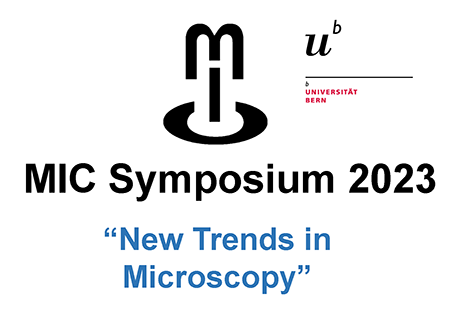
MIC Symposium 2023
The MIC Symposium was held in 2023 on November 17th with the theme “New Trends in Microscopy”.
The meeting was kicked off with a welcome address by Aurel Perren, the Deputy Dean of the Medical Faculty, and the exciting announcement by Britta Engelhardt, the president of the MIC, that the MIC was recently recognized as one of the five "official" core facilities of the University of Bern.
We moved on to the first session of our scientific program where we were introduced to methods by which single cells can be visualized in their large global context. Gail McConnell from the University of Strathclyde, UK presented optical imaging with the mesolens, a giant microscope objective that allows full 3D recording of many thousands of cells and amazed us by 3-D printing the lenses they use in their system. Thomas Nevian, Physiology, UniBern followed with his cutting edge work on how in vivo miniscopes can image single cell activity of neuronal cells in the brain of freely moving animals.
The second session took a bit of a turn to introduce how various ‘’omic’ technologies can be combined with imaging to provide spatial context of large proteomic, transcriptomic and metabolomic data sets. The session began with a talk by Hannah Williams, Institute of Tissue Medicine and Pathology (IGMP), UniBern who is coordinating the local Spatial Omics Consortium (SPOC, https://spoc-bern.squarespace.com) for those interested in the techniques and the latest developments in spatial omics. Joel Zindel, Visceral Surgery, UniBern presented his research on how he used spatial transcriptomics to identify new factors in abdominal adhesion formation. The session was concluded with a talk by from Daniel Schulz, Dept of Quantitative Biomedicine, UniZürich, in which he presented how imaging mass cytometry can be used to characterize protein and RNA in the tumor microenvironment and how this is used to compare 1000s of patient samples within the “Integrated iMMUnoprofiling of large adaptive CANcer patient cohorts” (IMMUcan) Consortium.
In our final session we had fascinating talks on new advances in super resolution and expansion microscopy. Paul Guichard, UniGeneva introduced us to the possibility of using expansion microscopy to increase the resolution of the molecular architectures by enlarging cells in a polymer matrix. Subcellular resolution imaging can then be achieved with conventional diffraction limited microscopes. The meeting was closed with an outstanding talk from Stefan Hell, Max-Planck-Institute, Germany in which he shared the history of developing super resolution microscopy that awarded him a Nobel prize and how he is continuing to develop the MINFLUX super-resolution microscope.
This year’s MIC symposium was a success due to the excellent speakers, and also from the excellent preparation by the MIC administration and our student helpers. And last, but not least due to the generous support we received from our industrial sponsors. The event was fully booked weeks in advance with over 200 registrations.
The scientific committee, Kerry Woods, Deborah Stroka, Thomas Nevian hope all attendees enjoyed the program and got an insight into new microscopy technologies that enable a more detailed look into the complexity of tissues and cells. Find more impressions here.
On Thursday, November 23, 2023, Prof. Konstantin Yenkoyan, Vice-Rector for Science and Head of Neuroscience Laboratory of Yerevan State Medical University after Mkhitar Heratsi (YSMU), Armenia, visited representatives of the Microscopy Imaging Center of the University of Bern. Konstantin is also the coordinator of the first-time-ever successful Horizon 2020 project at YSMU and the scientist-in-chief of the "COBRAIN" Scientific-Educational Center for Fundamental Brain Research (https://cobrainarmenia.org/; https://ysmu.am/en/research/mijazgayin-gitakan-tsragrer/cobrain/). This educational center aims at enhancing the research and innovation capacity of YSMU by strengthening the field of brain research with a special focus on autism and Alzheimer’s disease. The center offers a new model of research-based education and thus develops a new culture in higher education.
The visit of Prof. Yenkoyan to Switzerland was organized within the framework of the "Brain Visualization Laboratory" grant project, which is implemented in cooperation with "Center for Education Projects" PIU of the Ministry of Education, Science, Culture and Sports of the Republic of Armenia and the World Bank. In lively discussions with MIC committee members, Konstantin presented the main goal of the project, which is to establish a brain visualization laboratory by combining state-of-the-art microscopy, cellular computing, augmented and virtual reality, enabling comprehensive visualization of the brain’s cellular engineering. Other points of discussion included the research directions of the "COBRAIN" center and opportunities for collaboration between researchers in Bern and Yerevan.
Through this visit, Prof. Yenkoyan also received direct input on how a research-oriented bioimaging infrastructure can be organized, managed and operated. In addition to Bern, Konstantin Yenkoyan also visited imaging facilities in Zurich, Basel and Geneva.
SNSF Scientific Image Competition 2024
The SNSF Scientific Image Competition encourages researchers working in Switzerland to present their works to the public and the media. Photographs, images and videos will be rated in terms of their aesthetic quality and their ability to inspire and amaze, to convey or illustrate knowledge, to tell a human story or to let us discover a new universe. Find more information here.
All scientists working at a research institution in Switzerland are eligible to participate. The works must have been produced less than 12 months before the deadline for submitting entries.
The competition is held annually. An international jury will meet at the beginning of the year and award a CHF 1,000 prize in each category for the winning entry, as well as CHF 250 for each distinction. The award-winning works are announced in April or May, displayed in an exhibition at the Biel/Bienne Festival of Photography and made available to the public and the media, as well as to scientific institutions.
The competition has multiple aims: to highlight the growing role of images in scientific research, to reveal how scientific work is conducted and to give a face to the researchers conducting it. The competition also aims to encourage the media to use more images in their science coverage and make them accessible to the public through exhibitions.
Submission form.
Open Call for services from European research infrastructures, including Euro-BioImaging ERIC, for projects in the field of cancer research
The Horizon Europe provides a funding opportunity within the funded canSERV project.
This call offers financial support for services provided by European research infrastructures, including Euro-BioImaging ERIC, for projects in the field of cancer research. The deadline for submitting applications is January 4, 2024. For more information, please click here:
BioVisionCenter
Recently the BioVisionCenter was founded, a co-foundation by the Friedrich Miescher Institute and the University of Zurich, dedicated to all activities related to computer vision and advanced processing of complex bioimage datasets.
To celebrate the launch and foster collaboration within the scientific community, they are organizing the BioVisionCenter Symposium on the 9th and 10th of November 2023. This symposium will bring together renowned experts in the field of bioimage analysis who will share their insights, present cutting-edge research, and discuss emerging trends. It will be an excellent opportunity for researchers, students, and professionals to network, exchange ideas, and explore potential collaborations. Find out more about the symposium and sign up here.
MIC Symposium 2023 - Invitation
We are pleased to invite you to our annual MIC Symposium taking place on November 17, 2023 at the UniS in Bern.
This year’s chosen topic of the MIC Symposium is “New Trends in Microscopy”. A wide range of topics on novel and unconventional microscopy techniques will be covered by speakers from different countries as well as by speakers from the University of Bern and from industry. Don't miss the opportunity to learn about the latest trends in microscopy and to network with participants and speakers from all over Europe. The Scientific Committee formed by Kerry Woods, Deborah Stroka and Thomas Nevian, all University of Bern, will guide you through the day.
Registration is mandatory and places are limited. So register soon.
We look forward to meeting you at the MIC Symposium 2023 and are pleased to present a wide range of speakers and a day filled with exciting presentations, discussions and exchanges with all of you.
Swiss Microscopy Core-Facility Day 2023
11th Swiss microscopy core facility day this year took place on September 14, 2023 in Zurich and was organised by ScopeM, an imaging platform of ETH Zurich. About 50 members of Swiss imaging facilities and representatives of microscopy companies convened for a day to discuss the questions related to recent microscopy developments and different aspects of microscopy faculty operation. In the morning talk, Matthia Karreman from DKFZ in Heidelberg, presented her research on imaging preclinical models of brain metastases with LM-uCT-EM corelative approaches. In the afternoon, sessions the number of facility and scientific speakers covered the subjects of super resolution and spatial omics, particularly how these new technologies can be implemented in an imaging facility. At the end of the meeting, participants were able to tour the ScopeM and see the most interesting systems there and learn about projects performed on them. The meeting finished with an informal apéro where the participants had a chance to network with each other and talk to speakers and industry delegates. Yury Belyaev represented MIC in this meeting. Next year the Swiss core facility day will be taking place in Geneva or Lausanne.
Industry Contest for young scientists
You are a young scientist, postdoc or junior professor in Germany, Austria, Switzerland or the Netherlands? You are working on an exciting cell biology project but do not have access to confocal high content imaging? The imaging part is missing for a step forward or your next publication?
Submit your imaging assay idea in the contest and win access to high quality results.
COMULIS Training School "Imaging Across Scales"
The five-day COMULIS Training School "Imaging Across Scales" took place at the University of Bern on September 4-8, 2023. Organized by Ruslan Hlushchuk, Benoît Zuber (Institute of Anatomy) and Yury Belyaev (Microscopy Imaging Center) this event offered a balanced mix of theory and hands-on experience for 9 students from 4 European countries.
The event started with a keynote talk by Stephan Handschuh of Vet-Med University of Wien on correlative workflow. Days 2 and 3 showcased diverse perspectives with speakers such as Ruslan Hlushchuk on uAngio CT, David Haberthür on uCT analysis, Paolo Ronchi from EMBL Heidelberg on X-ray tomography, and Benoît Zuber on SBF SEM. Industry views were presented by Phil Salmon from Bruker microCT and Emine Korkmaz from ThermoFisher Scientific. Joana Delgado Martins from University of Zurich discussed big data storage and handling.
Participants, divided into four rotating groups, took part in hands-on sessions covering uAngioCT sample preparation, uCT and EM imaging workflows and image processing. These sessions enabled a practical application of the topics discussed in the lectures and enabled participants a possibility of one-to-one communication with imaging experts.
The final day included group presentations and a feedback session, rounding off a week of comprehensive learning and networking, which also included a welcome dinner and a city tour. Overall, the program successfully fused expert talks, practical training, and networking activities.
The training school was supported by the Institute of Anatomy, Microscopy Imaging Center, and COMULISglobe - Chan Zuckerberg Initiative.
Summer Series 2023 from the Dubochet Center of Imaging
The Dubochet Center of Imaging (DCI) summer 2023 seminar series is an excellent opportunity to hear the latest in cryo-EM and cryo-ET from leading researchers in the field.
The next dates and speakers will be:
- Arjen Jakobi (TU Delft, NL) on 01.09.23, 12:00, at EPFL Lausanne (Cubotron, BSP231)
- David Barford (MRC LMB Cambridge, UK) on 15.09.23, 11:00, at Uni Geneva
- Florian Schur (ISTA Klosterneuburg, Austria) on 22.09.23, 13:15, at Uni Bern (Ewald Weibel Auditorium, Institute of Anatomy)
Please contact us if you would like to have any further information.
CEM Summer School 2023
From July 6th to 7th, 2023, the annual summer school of the PhD program Cutting Edge Microscopy (CEM) took place at the Seehotel in Bönigen, Switzerland. Bönigen is a beautiful village on Lake Brienz. All students gave scientific presentations on their projects, with a focus on the microscopy applied and the results obtained. Since the students’ projects cover very different topics and a broad variety of microscopy, a new structure was tried: For each session, groups of 2 to 4 students were formed depending on the type of microscopy used or their scientific project. To introduce and explain the microscopy, the students were encouraged to make joint presentations. This format encouraged a more lively atmosphere and was greatly appreciated by all participants. The even livelier review of the scientific part of the summer school took the form of a quiz in which each student contributed to a question pool covering all presentations. Advice on how to implement microscopy in the projects was provided by the MIC microscopy specialists Fabian Blank and Yury Belyaev, the MIC bioimaging specialist Guillaume Witz and the CEM PhD program co-directors Benoît Zuber and Steven Proulx. Ruth Lyck, the CEM PhD program coordinator, was responsible for the quiz and hotel organization. In addition to the scientific program, a discussion on upcoming activities of the CEM PhD program took place. As the new CEM students’ representatives Adrian Madarasz and Marwa Mangattu Parambil were elected. During their term from August 2023 until July 2024, Adrian will join the MIC committee as the students’ representative. Another much appreciated activity was the visit of Leica Microsystems representatives Annette Rinck, CEO, and Michael Weuffen, Service manager. The beautiful landscape, the pleasant accommodation and free time to swim in Lake Brienz or go jogging contributed to a perfect learning atmosphere.
ELMI 2023
The 22nd International European Light Microscopy Initiative Meeting place on June 6-9, 2023 in Noordwijkerhout, The Netherlands. More than 400 participants from Europe and the whole world discussed recent advances in light microscopy aimed at a life sciences audience.
During the first day of the meeting, in a traditional core facility session, the representatives of the imaging facilities discussed the topics of common interest such as cyber security, facility equipment maintenance and tools, staff development, data management. During the following two and a half days of the conference the leading scientists in the field presented and discussed with delegates the latest achievements in microscopy. The topics of this year were: Functional imaging, new technologies, data management & analysis, intravital & light sheet, CLEM, and high content. Additionally, more than 50 leading companies active in microscopy instrumentation and related fields introduced latest cutting-edge technical developments in informative workshops and booth presentations. As usual, ELMI provided an excellent opportunity for networking with colleagues from Industry and Academia.
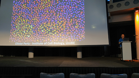
MIC was well represented by three MIC members. Olivier Pertz (Institute of Cell Biology), Ruslan Hlushchuk (Institute of Anatomy) and Yury Belyaev (MIC) attended the meeting. Olivier gave an invited talk ‘’Spatio-temporal interrogation of signalling networks using biosensor imaging and optogenetics’’ covering the latest work in his lab. Ruslan in his talk ‘’Towards high accuracy µCT-LM-EM correlative workflow with fluorescent µCT contrast’’ presented along with the research of his group also a collaborative project with Yury Belyaev and colleagues from the University of Fribourg (Felix Meyenhofer, Boris Egger) and the University of Porto (Paula Sampaio, Ines Alencastre and Maria Azevedo).
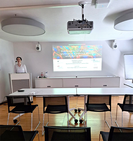
Congratulations to successful PhD Defense Anastasia Milusev
On July 6, 2023, Anastasia Milusev, student at the PhD program Cutting Edge Microscopy (CEM) under the supervision of Prof. Dr. Robert Rieben and Dr. Nicoletta Sorvillo has successfully defended her PhD thesis with the title "Distinct arterial and venous glycocalyx dynamics impact endothelial function".
Abstract: The endothelial glycocalyx, a layer of sugars and proteins on the endothelial cell surface, is crucial for maintaining vascular homeostasis. The glycocalyx is damaged in different inflammatory conditions, leading to a pro-inflammatory and procoagulant endothelial cell phenotype. Using confocal microscopy and live cell imaging, the glycocalyx dynamics of arterial and venous endothelial cells in different inflammatory conditions were investigated in vitro in a 3D microfluidic system.
MIC Research Day 2023
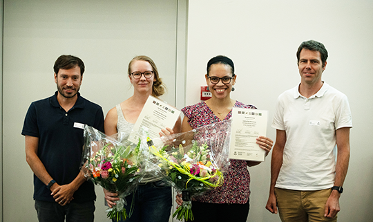
On June 28, 2023, more than 120 participants from the University of Bern, Fribourg, Geneva, and industry representatives attended the traditional MIC research day, that took place at UniS. The well-balanced program of the event featured MIC users from all three Faculties of the MIC (Vetsuisse, Natural sciences and Medicine) as well as one researcher from the University of Fribourg and one industry speaker.
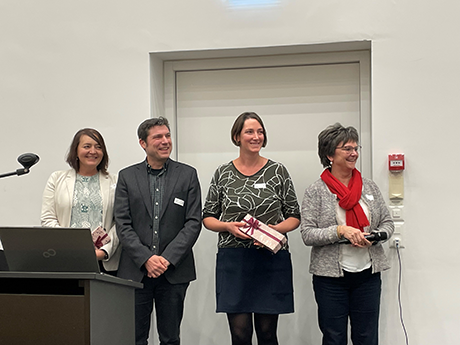
Nine scientists presented their research projects related to microscopy. Among them, Ora Hazak from the University of Fribourg introduced her studies of plant receptor-ligand signalling using fluorescence lifetime imaging. An industry representative, Nikolaos Droseros from GMP, informed the audience about the latest developments of lasers for multiphoton microscopy. Finally, two students who successfully graduated from the PhD program Cutting Edge Microscopy (CEM) received their certificates.
As usual, this event provided plenty of opportunities for networking among the imaging community at the University of Bern. We thank all CEM students for their help in the organization, all participants for their enthusiasm for microscopy and event sponsors for their generous financial support.
Inauguration Symposium for new electron microscopes at the Institute of Anatomy on July 5, 2023
The inauguration symposium to celebrate acquisition of the Titan Krios G4 transmission cryo-electron microscope and the Aquilos 2 cryo-focused ion beam milling scanning electron microscope takes place in the Ewald R Weibel Auditorium (Room No. A224) at the Institute of Anatomy, Bühlstrasse 26, 3012 Bern, on July 5, 2023. Prof. Werner Kühlbrandt of the Max-Planck Institute of Biophysics in Frankfurt will be sharing his insights in a scientific talk entitled “High-resolution cryoEM of mitochondrial membrane proteins”. Registration via e-mail to office.ana@unibe.ch.
Invitation to the MIC Research Day 2023
We are pleased to invite you to our annual MIC Research Day on June 28, 2023.
The MIC Research Day shows a selection of research highlights based on microscopic techniques that have been realized by scientists from the University of Bern. Junior and senior researchers who are specialists in microscopy present data on their exciting research projects, which were achieved using state-of-the-art microscopy. With this event, the MIC brings together scientists using microscopy with other researchers who may wish to use the same instrument or a similar technique.
We are happy to present a day full of interesting topics and discussions and we are honoured to welcome great speakers who will share their expertise on microscopy in their various research areas.
We are looking forward to welcome you in June.
Find all information and registration here.
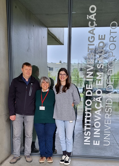
Short term scientific mission in Porto, Portugal, by Dr. Yury Belyaev
In April 2023, Dr. Yury Belyaev, light microscopy manager of the Microscopy Imaging Center of the University of Bern, visited the Advanced Light Microscopy (ALM) and the Bioimaging scientific platforms at i3S - Instituto de Investigação e Inovação em Saúde, the University of Porto, Portugal. For this 5-day short-term scientific mission (STSM), Yury Belyaev received financial support from the COST (European Cooperation in Science and Technology) Innovator Grant COMULIS (Correlated Multimodal Imaging in Life Sciences) MultEMPlex IG17121.
The aim of STSM was to show a feasibility of implementation of super-resolution and lifetime microscopy into the existing micro computer tomography(µCT)/light microscopy (LM) correlative workflows at the University of Bern. The samples of mouse kidney from the µCT group of PD Dr. Ruslan Hlushchuk, Institute of Anatomy, University of Bern, were investigated using the Leica Stellaris 8 FALCON (FAst Lifetime CONtrast) STED (STimulated Emission depletion) confocal microscope. Different sample preparation protocols suitable for high-resolution STED imaging have been also tested.
The obtained results allow higher correlation precision with additional tissue specificity and possibility of label-free imaging in existing multimodal correlative workflows and open the possibility to develop it further in the direction of electron microscopy. The scientific outcome of STSM will be presented in one of the upcoming meetings on correlative multimode imaging. Finally, the visit has contributed to the development of scientific collaboration between Portugal and Switzerland.
The Grantee would like to thank COST Action for financial support and the members of the ALM and Bioimaging scientific platforms at the i3S institute for their hospitality, friendly atmosphere, and excellent scientific input during this STSM.
PhD Defense L. van Os
Lisette van Os, student at the PhD program Cutting Edge Microscopy (CEM) under the supervision of Prof. Dr. Olivier Guenat, will give her thesis defense on April 27, 2023.
Title: Immune cell migration during infection in organs-on-chip
Abstract: Organ-on-chip models aim at replacing traditional animal experiments in the research and drug development pipeline. Current organ-on-chip models can replicate the functional unit of an organ, but often do not include an immune component. In this project, an organ-on-chip model of lung infection was developed, to study immune cell migration during the acute infection phase. Live microscopy was carried out to follow the migration pattern of immune cells under different (inflammatory) conditions. Immune cell migration during lung inflammation could be visualized in real time.
PhD Defense S. Mutlu
Seyran Mutlu student at the PhD program Cutting Edge Microscopy (CEM) under the supervision of PD Dr. Fabian Blank and PD Dr. med. Amiq Gazdhar will give her PhD Defense on April 26, 2023.
Title: Adoptive transfer of HGF overexpressing T cells as potential therapeutic approach for bleomycin injured lung mouse model
Abstract: Idiopathic pulmonary fibrosis (IPF) is a lethal disease emerging from aberrant wound healing mechanisms disturbing the alveolar epithelial (AE) integrity. The immune system (IS) is known to be involved too. Hepatocyte growth factor (HGF) showed antifibrotic properties. We aim to study if adoptive transfer of HGF modified T cells (HGF-T cell treatment) in bleomycin injured mice (BLM model) may promote balancing of the pulmonary IS and repair of the AE.
We established a model of precision cut lung slices, which were analyzed by confocal microscopy in order to observed instillated T cells after treatment in the murine lung. Live-cell imaging was operated on co-cultures using the Incucyte system to see if HGF transfected T cells were interacting with myofibroblasts.
Congratulations to successful PhD Defense Daniëlle Verschoor
On February 10, 2023 Daniëlle Verschoor, student of the Cutting Edge Microscopy (CEM) program under the supervision of Prof. Stephan von Gunten, has successfully defended her PhD thesis with the title: “Granulocytes, fragile but resistant. Unraveling the treatment resistance of granulocytes.”
Her PhD project focused on the treatment resistant of inflammatory granulocyte, both neutrophils and eosinophils. Freshly isolated human peripheral granulocytes were stimulated with pro-inflammatory cytokines as GM-CSF and/or IL-5 followed by treatments as intravenous immunoglobulins, anti-siglec-7/9 or glucocorticoids to investigate their cell survival or death and to investigate possible mechanism involved in this response. In her project microscopy was used to characterize subsets of cells and analyze/visualize cell death. This all to unravel the mechanism responsible for their resistance to these treatments.
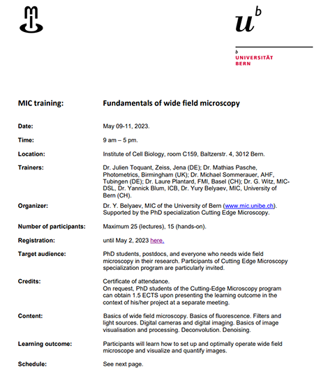
Fundamentals of wide field microscopy
The MIC Training "Fundamentals of wide field microscopy" will take place on May 09 - 11, 2023. Content of the course will be: Basics of wide field microscopy. Basics of fluorescence. Filters and light sources. Digital cameras and digital imaging. Basics of image visualisation and processing, deconvolution and denoising.
On request, PhD students of the Cutting-Edge Microscopy program can obtain 1.5 ECTS upon presenting the learning outcome in the context of their project at a presentation event.
Registration closes on May 2, 2023.
Detailed information and the registration form are to be found on ILIAS.
Demonstration Events
The InCell Analyser (INCA) high-throughput microscope at the Vetsuisse Faculty has to be replaced soon. To learn about the performance of instruments on the market demonstration events will be held for the Molecular Device (Nano/Micro) instrument on March 27-30 and for the Cytation 5 and 10 instruments on April 18-19, 2023. Please contact Prof. Kässmeyer, Dr. Kerry Woods or Dr. Helena Röss if you are interested in joining these demonstration events.

MIC Workshop: Performance Test in Light Microscopy
In collaboration with the Friedrich Miescher Institute for Biomedical Research in Basel, the MIC will offer the course “Performance Test in Light Microscopy”. It will take place on April 26-27, 2023. The course will present some basic performance tests to diagnose issues and benchmark performance of light microscopes. Please find all further information on the flyer below.
In case you are interested, please send your application to: yury.belyaev@mic.unibe.ch
Application deadline: March 10, 2023
CEM Study Trip 2023
In February 2023, a study trip took the PhD students of the PhD program Cutting Edge Microscopy (CEM) to Freiburg i.B., Germany. Here, the students visited the Life Imaging Center (LIC) of the University of Freiburg. The institute is headed by Dr. Roland Nitschke, who warmly welcomed the students and led them through the 2-day program together with his team. A relaxing tour through the beautiful city center of Freiburg i.B. completed the program. Read more about the study trip of the CEM PhD students to Freiburg in the pdf file linked below.
Advanced Imaging Course - UniFR
The Microscopy Imaging Center (MIC) collaborates with the Bioimage Core Facility of the Department of Biology and the Section of Medicine of the University of Fribourg. The course Advanced Imaging - Super-resolution fluorescence microscopy is open for PhD students of the Cutting Edge Microscopy specialization program.
Fluorescence microscopy has become the preferred imaging tool for biological systems due to its capability to visualize specifically the target biomolecules under conditions compatible with life, like room temperature, liquid environment and irradiation with visible light. Fluorescence microscopy also provides very high sensitivity down to the detection of single molecules. As drawback, as it happens with any a far-field optical technique, the spatial resolution is limited by the wavelength of light to a few hundreds of nanometres (the so-called diffraction limit). Remarkably, in the mid 2000s, a series of imaging methods using fluorescence readout were developed that deliver images with resolution beyond the diffraction limit. These methods, called super-resolution fluorescence microscopy or far-field fluorescence nanoscopy and whose pioneers were awarded with the Nobel Prize in Chemistry in 2014, have revolutionized biological imaging and continued to be developed until the present day.
Detailed information are to be found on ILIAS here. KSL: 482916
PhD Defense M. Golomingi
Murielle Koni-Kepo Golomingi student at the PhD program Cutting Edge Microscopy (CEM) under the supervision of Prof. Dr. Verena Schröder, will give her PhD Defense on March 15, 2023 at 14.00 at the ARTORG Center for Biomedical Engineering Research.
Title: “Interactions between the complement system and blood coagulation: a potential role of complement components and activation during haemostasis"
Haemostasis is a crucial process by which the body stops bleeding. It is achieved by the formation of a platelet plug, which is strengthened by the formation of a fibrin mech through the coagulation cascade. In case of pathological clotting, for instance in arterial thrombosis, the interactions of the complement system and the coagulation cascades is known to aggravate the development of the pathology. Whether those interactions play a relevant role during non-pathological haemostasis is however not yet completely understood. The aim of this study was to investigate the potential role of complement components and activation during haemostasis.
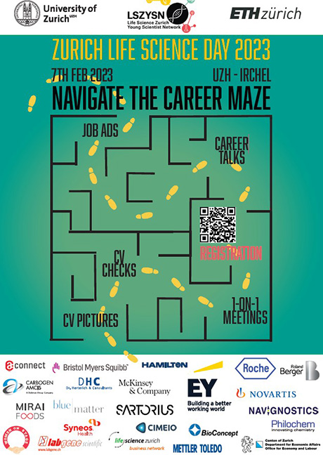
It is time to navigate your career maze!
The Life Science Zürich Young Scientist Network (LSZYSN) invites you to the Zurich Life Science Day, on the 7th of February 2023 at Irchel Campus. This is a day-long career fair and networking event with the most talented life scientists and companies.
The LSZYSN is a non-profit organization composed of PhD students and Post-Doctoral researchers from ETH and University Zurich. Our goal is to connect life scientists with their career opportunities beyond academia.
Over the last 12 years, the ZLSD has grown into the largest job fair for life scientists in Switzerland, hosting more than 700 participants and 100 company representatives on site at the Irchel Campus of the University of Zurich. Due to the Covid-19 situation we went virtual the last 2 years and hosted over 2000 participants from all over the globe, who connected with company representatives over informal 1-1 meetings, and numerous chatting options. However, ZLSD is back on campus and you get to do all networking in person.
Muldimodal Imaging
On February 2, 2023 the AGORA Cancer Research Center invites you to the workshop titled "Multimodal Imaging in life sciences and cancer research". Keynote speaker is Ralph Weissleder, Harvard/ MGH, USA. More details and registration on the pdf below.
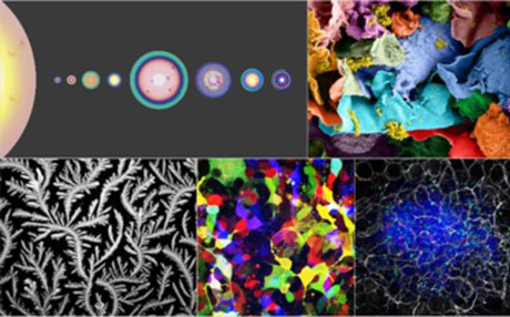
SNSF Scientific Image Competition
Get your cameras and give Swiss research a face!
The SNSF Scientific Image Competition encourages researchers working in Switzerland to present their works to the public and the media. Photographs, images and videos will be rated in terms of their aesthetic quality and their ability to inspire and amaze, to convey or illustrate knowledge, to tell a human story or to let us discover a new universe.
Submission deadline is January 31, 2022. Learn mor here.
First protocol for Monitoring the point spread function
MIC congratulates Dr. Yury Belyaev on his co-authorship in a new publication on the use of the point spread function for quality control of confocal microscopes. The paper can be downloaded here.
Benchtop High-Speed Confocal Microscope
Yury Belyaev (MIC) and Fabian Blank (DBMR) invite you to this demonstration event organized in collaboration with Witec AG. More details and registration on the flyer below.
Congratulation to successful PhD Defense
On September 20, 2022 Raphaela Seeger, student of the PhD program Cutting Edge Microscopy (CEM) has successfully defended her PhD thesis entitled "Investigating synaptic vesicle exocytosis by structural studies of rat synaptosomes".
During this project synapses were isolated and treated with an exocytosis triggering solution prior to vitrification by plunge freezing to achieve a temporal resolution and investigate the morphofunctional changes in the synapses' architecture via cryo-electron tomography. In parallel, tomograms of synapses with a mutated exocytosis-relevant-molecule (SNAP25) from collaborators (University of Copenhagen) were evaluated and compared with the results of the time-resolved exocytosis events. Further, a deep-learning software to automate the segmentation of synaptic vesicles was created in a collaboration with another PhD student from the Zuber lab.
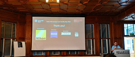
Swiss Microscopy Core Facility Day 2022
The 10th Swiss Microscopy Core Facility Day took place this year on September 15 - 16, 2022 and was organised by Pascal Lorentz from the Department of Biomedicine at the University of Basel. To celebrate the 10th anniversary, this normally one-day event, was organised as a two-day retreat in Engelberg. About 60 members of Swiss microscopy facilities and representative of different microscopy companies convened for informative presentations and intensive discussion on questions related to various aspects of microscopy faculty operation. The focus of this year’s meeting was the well-being of the facility staff.
On the first day, the leading microscopy companies shared their experiences in supporting good working atmosphere among employees. Additionally, Christel Genoud from the University of Lausanne presented the results of a facility survey on the working conditions of microscopy facility employees. On the second day, after Zeiss’ presentation of Zeiss on the latest developments in artificial intelligence (AI) software and Evident’s brief overview of corporate wellbeing issues, the participants were able to participate in variouse workshops. All workshops were led by professional coaches and gave an insight into modern psychological methods tackling questions of mental health, self-reflection and conflict management.
As usual, the meeting offered many opportunities for networking and exchange of experiences during its social program. Yury Belyaev represented the Microscopy Imaging Center (MIC) at this meeting. Next year the Swiss Core Facility Day will take place at ETH Zurich organised by ScopeM.
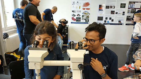
MIC at "Nacht der Forschung 2022"
The Nacht der Forschung at the University of Bern took place on September 10, 2022. The room of the microscopy imaging center (MIC), UniS A015, was decorated with various posters about the MIC and about microscopy at our University. Activities included "Microscopy for everyone" on 6 teaching microscopes and an excursion into virtual reality, which beamed the guest into the brain of a mouse or into a fish head. In room A-126 of UniS, microscopists offered lectures on exciting topics. A total of around 30 helpers from the MIC committee, friends of the MIC and students of the PhD program Cutting Edge Microscopy headed by Fabian Blank, Ruslan Hlushchuk and Ruth Lyck were on duty in 3 shifts from 4:00 p.m. to midnight. Further activities of MIC committee members were offered in the University’s main building by Stefan Tschanz and Benoît Zuber on behalf of the Institute of Anatomy. All activities were eagerly attended. Interested conversations and happy children's faces testified to the success of our offers. The MIC team says a big "THANK YOU" to all the helpers, without whom the MIC activities would not have been possible.
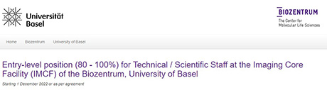
Scanning System VS200 and All-in-One Microscope APX100
This workshop, organized in collaboration with Yury Belyaev (MIC) and Fabian Blank (DBMR), will take place on October 5, 2022 at the Department for BioMedical Research, Bern. For further details please see the flyer below.
Entry-level position at the Imaging Core Facility (IMCF)
The Imaging Core Facility (IMCF) of the Biozentrum of the University of Basel is offering a position to a young scientist to enter the field of modern high-end light microscopy. More details see here.
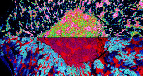
Translational molecular imaging: Using a mass spectrometer as a microscope
The Department for BioMedical Research (DBMR) at the University of Bern is hosting Prof. Dr. Ron M.A. Heeren from the Molecular Imaging Institute, Maastricht University at the DBMR Research Conference.
The conference is taking place on October 31, 2022 at the Langhans Auditorium. More information here.
PhD midterm presentation by MD Adrian Madarasz
On December 16, 2022 at 2.00 pm. Title: “Circulation and clearance of cerebrospinal and brain interstitial fluid in murine models of hemorrhagic stroke". Location: Theodor Kocher Institute, University of Bern.
MD Adrian Madarasz is enrolled in the PhD program Cutting Edge Microscopy and conducts his thesis under the supervision of Dr. Steven Proulx at the Theodor Kocher Institute
Abstract of his presentation: Hemorrhagic stroke is associated with high disability and mortality rates. Most therapeutic approaches lack broad agreement, especially for the reduction of brain edema. By inducing disease models for intracerebral and subarachnoid hemorrhage in mice followed by in vivo imaging with near-infrared tracers and labels, we assess changes in brain interstitial and cerebrospinal fluid circulation and investigate potential efflux pathways for fluids and cells from the subarachnoid space.
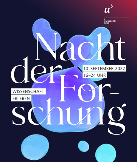
Meet the MIC at the "Nacht der Forschung"
The MIC team invites the public to inspect fascinating specimen through a microscope, to see posters about the MIC and some microscopy examples done at the University of Bern and to enjoy 3D images in virtual reality in room A015. In addition, MIC experts will held presentations at 5.00, 6.00, 8.00, 9.00 and 10.00 pm in room A-126.
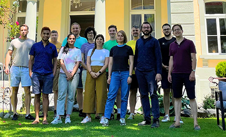
Workshop on Bioimage Processing with Python
From July 11 to 15, 2022, the 1st MIC Workshop on Bioimage Processing with Python, led by MIC team member Dr. Guillaume Witz, took place in the Bernese Oberland. A mix of eight participants from the University of Bern, three from European universities and one from a private company learned how to develop Python-based bioimage processing workflows using cutting edge tools like cellpose, napari etc. Participants also had the opportunity to work on their own projects and to discuss them with Guillaume Witz and with each other. It was a great experience to see how concepts and tools just taught were applied live to those projects, and how participants made massive progress in such a short time! During breaks and evening time the beautiful park of Sherlock Holme's favourite hotel in Meiringen was a wonderful stage for many more informal discussions on science, microscopy, biology etc. made especially interesting by the participants' diverse backgrounds. Finally, participants also enjoyed the many hiking and excursion possibilities available in Meiringen to recover from an intensive learning program. Inspired by this success, MIC will organize the 2ndMIC Workshop on Bioimage Processing in 2023. Stay tuned and visit our website for announcements!
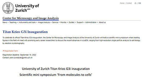
CryoEM symposium - UHZ, Center for Micoscopy and Image Analysis
The UZH Center for Microscopy and Image Analysis invites to a mini-Symposium dedicated to the inauguration of the Titan Krios G3i on Monday, 3rd October 2022 at the Irchel Campus, University of Zürich. The Krios series represents cryo-transmission electron microscopes (TEMs) with atomic resolution. Therefore, this symposium could be helpful for you if you are dealing with TEM in your research projects.
Congratulations to an important research achievement!
Congratulations to Dr. Coralie Dessauges and the research group of Professor Olivier Pertz, Institute of Cell Biology, Faculty of Science, University of Bern, on the publication of their project "Optogenetic actuator – ERK biosensor circuits identify MAPK network nodes that shape ERK dynamics" in the EMBOpress journal Molecular Systems Biology in June 2022, downloadable at https://doi.org/10.15252/msb.202110670. In this work, Coralie and co-workers built circuits consisting of an optogenetic actuator to activate MAPK signaling with blue light and an ERK biosensor to measure single-cell ERK dynamics. The inducing light pulse is automatically provided by the microscope, allowing the simultaneous study of dynamic ERK responses in thousands of cells. In summary, this project improves our understanding on the MAPK signaling interactome.
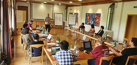
CEM Summer School 2022
On June 30th and July 1, 2022, the annual summer school of the PhD program Cutting Edge Microscopy (CEM) took place at the Hotel Appenberg in Zäziwil, Switzerland. On the first day, the CEM PhD students presented their scientific projects and specific microscopic applications. Advice on the implementation of microscopy into the projects was provided by the MIC microscopy specialists Fabian Blank and Yury Belyaev, the MIC bioimaging specialist Guillaume Witz and the CEM PhD program co-directors Benoît Zuber and Steven Proulx. Furthermore, a discussion on the next activities of the CEM PhD program took place and Javier Pareja and Adrian Madarasz were elected as CEM student representatives from August 2022 to July 2023. On the second day, the external trainer Michael Berndonner carried out a very intensive training course in scientific presentation techniques. Moderation skills were also included in this course as an important topic for scientific conferences. The coordinator of the CEM PhD program, Ruth Lyck, also attended the CEM summer school and took care of all administrative issues. The beautiful landscape, the pleasant accommodation and the Appenberg games activity contributed to a perfect learning atmosphere.
Enabling imaging across scales
The First EMBL Imaging Centre Symposium ‘’Enabling imaging across scales’’ took place on May 31, 2022 at EMBL Heidelberg, Germany. At this meeting, Dr. Yury Belyaev represented the MIC and here reports about it.
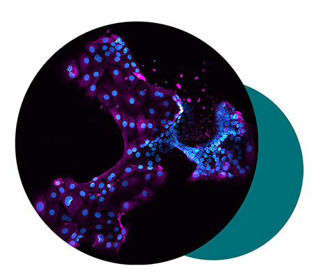
21st International European Light Microscopy Initiative Meeting (ELMI)
The 21st International European Light Microscopy Initiative Meeting (ELMI) took place on June 7-10, 2022 in Turku, Finland. At this meeting, Dr. Yury Belyaev represented the MIC and here reports about it.
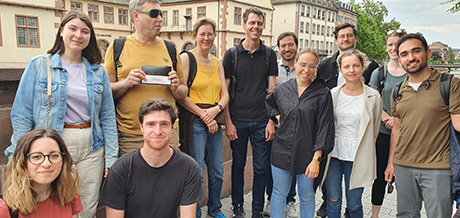
CEM Study Trip
In May 2022, a study trip took the PhD students of the PhD program Cutting Edge Microscopy to Strasbourg, France. Here, the students visited the Institute de génétique et biologie moléculaire et cellulaire (IGBMC). A relaxing tour through the beautiful center of Strasbourg completed the program. Read more about the workshops at the IGBMC on the pdf file below.
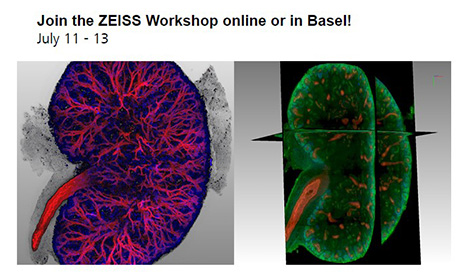
ZEISS Workshop online or in Basel
A Workshop dedicated to Tissue Clearing is jointly organized by The Imaging Core Facility (IMCF) of the Biocenter of the University of Basel and Zeiss on July 11 to 13, 2022
You will get the chance to explore this technology in different lectures in Basel or online. Specialists for ECI Clearing, Cubic Clearing and Smart Clearing will share their experiences. In the second part we will host a hands-on workshop, where you can carry out a selected clearing technique yourself. Further information here.
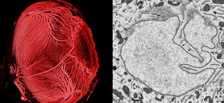
COMULIS training school "Imaging Across Scales"
29 August - 2 September 2022, Bern, Switzerland
The Microscopy Imaging Center and the Institute of Anatomy are organising a COMULIS training school ‘’Imaging across the scales’’ on 29 August – 2 September 2022 in Bern, Switzerland. This is a 5-days in-presence course with 16 participants. The talks and hands-on sessions will cover uCT-EM workflows. The course main topics are: uAngioCT, uCT and EM sample preparation, multimodal imaging, and image processing. Registstration here.
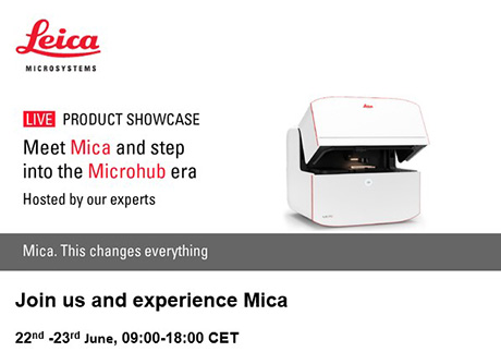
Mica microscope workshop at the Institute of Pathology
22nd -23rd June, 09:00-18:00 CET
It is our pleasure to invite you to book your personal demonstration of Mica - the world's first Microhub – hosted by the University of Bern at the Institute of Pathology. Discover true simultaneous 4-color widefield, confocal resolution and AI-supported analysis united in a sample-protecting incubator. All with a single push of a button!
Mica is a whole new way to do microscopy. Here’s why…
- Access for all
Automatic selection and optimization of imaging settings simplifies your experiment set-up and increases reproducibility - No constraints
Visualize 4 colors simultaneously in widefield and then switch to confocal without ever moving your sample. Select the right modality in real time - Radically simplified workflows
Intelligent automation and AI-supported analysis enables greater efficiency and a faster track to publication
Contact us to book a time for your live demonstration and have your questions answered.
Where: University of Bern, Institut für Pathologie, L570, Murtenstrasse 31, 3008 Bern
If you are not available during this time, please write an E-Mail.
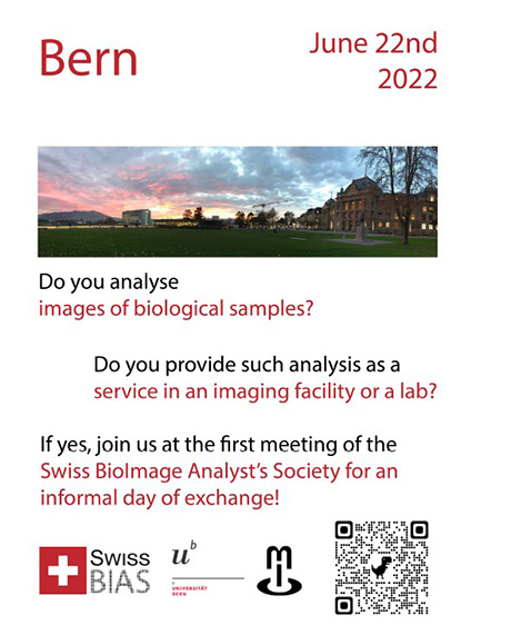
First meeting of the Swiss BioImage Analyst's Society
June 22, 2022
Do you analyse images of biological samples? Do you provide such analysis as a service in an imaging facilty or a lab?
If yes, register at the first meeting of the Swiss BioImage Analyst's Society for an informal day of exchange.
Swiss TriM Scope and Ultra Microscope User Meeting
Welcome to the Swiss User Meeting taking place on June 28, 2022. For program and registration please see flyer below.
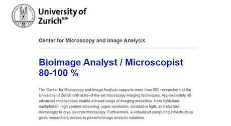
Open position as Analyst / Microscopist
The Center for Microscopy and Image Analysis at the University of Zurich offers a 4 year position. Further details see here.
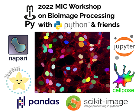
11 - 15 Jul 2022: MIC Workshop on Bioimage Processing
If you want to get started using the Python eco-system for your bioimaging projects this intensive one-week workshop is the right thing for you. In addition to course time, you will also have some time to work on your own projects, just getting started or continuing an existing one, and will be able to discuss them with other participants and the instructor. PhD students of the GCB profit from a reduced price. More details on the pdf below, registration here.
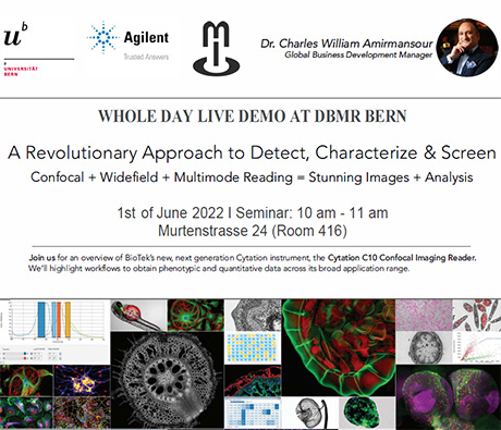
Whole day live demo at DBMR Bern
A Revolutionary Approach to Detect, Characterize & Screen
Confocal+ Widefield + Multimode Reading= Stunning Images+ Analysis
Join us for an overview of BioTek's new, next generation Cytation instrument, the Cytation C10 Confocal Imaging Reader.
We' ll highlight workflows to obtain phenotypic and quantitative data across its broad application range . Registration here.
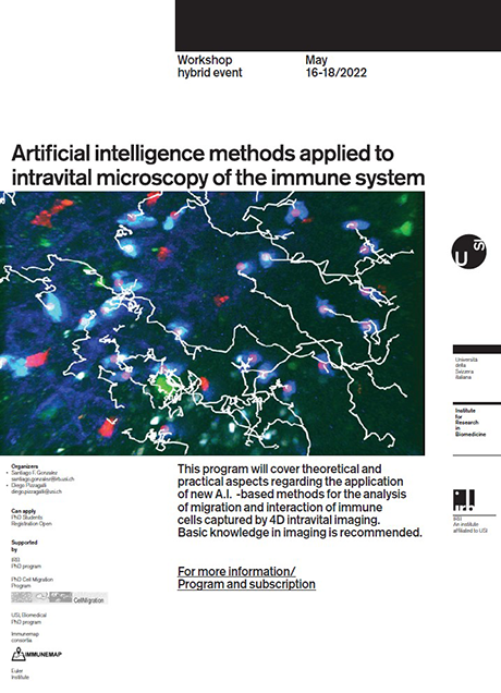
Artificial intelligence methods applied to intravital microscopy of the immune system
This hyprid workshop will cover theoretical and practical aspects regarding the application of new A.I. -based methods for the analysis
of migration and interaction of immune cells captured by 4D intravital imaging. Basic knowledge in imaging is recommended.
More information and registration here.
LS2 Prestige Webinar
MIC committee member Benoit Zuber, as part of the LS2 Biophysics section, invites you to the upcoming LS2 Prestige Webinar.
Topic: “CryoEM structures of amyloid filaments from human brain”
When: 30th March 2022, 16:00 – 17:00 (CET)
Please register here.
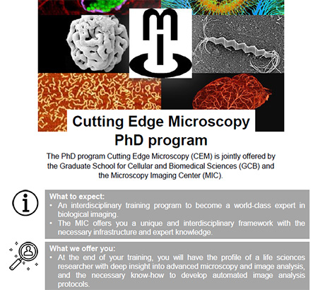
Join the Cutting Edge Microscopy PhD program
Interested in becoming a world-class expert in biological imaging?
The CEM PhD program currently has a few vacancies. If you are a PhD student of the GCB and you apply advanced microscopy techniques in your PHD thesis, we are looking for you. Please find more details on the flyer below and do not hesitate to contact us.
Midterm presentation
Irene Spera, student of the PhD program Cutting Edge Microscopy (CEM), will give her midterm presentation on March 9, 2022 at 11:00 online via zoom. Title: "Factors regulating the exchange of cerebrospinal fluid and interstitial fluid of the brain".
Our project investigates the perineural outflow pathways of cerebrospinal fluid (CSF) at the cribriform plate and focused on the elucidation of anatomical connections between the subarachnoid space (SAS) and the lymphatics at the exiting nerve routes. Furthermore, we want to define the anatomical spaces of the CSF flow on the brain surface and elucidate the relationship between CSF and the interstitial fluid (ISF) of the brain. To address these aims, we made use of transgenic reporter mice, fluorescence microscopy, and through skull near-infrared (NIR) in vivo imaging.
Please request acess to this online event via e-mail to Rahel Eyer.
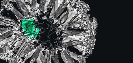
Join the Correlative Microscopy Symposium
March 13 - 16, 2022
The Organization Committee and the ZEISS Microscopy Team are delighted to invite you to the 6th Joint Meeting Workshop and Symposium “From 3D Light to 3D Electron Microscopy” taking place from March 13th to 16th 2022 in cooperation with the EMBL in Heidelberg, the VIB in Ghent and the Francis Crick Institute in London.
Many prominent scientists will share their groundbreaking work on correlative microscopy in 3D and volume EM. You can also look forward to practical workshops on
- correlative workflows
- volume EM methods
- sample preparation methods
and on a panel discussion of the international networking happening to promote these techniques.
This meeting will inspire you and open up the possibilities for forming new collaborative efforts to use these powerful imaging techniques to better understand the nano-world inside cells and tissues.
Please register here.
DCI Symposium 2022
The Dubochet Centre for Imaging (DCI) Symposium 2022 will take place in Lausanne and in Geneva June 8 to 10. More details and registration here.
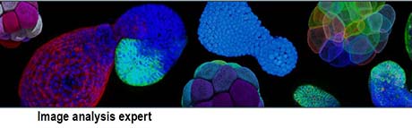
Image analysis expert
The Friedrich Miescher Institute für Biomedical Research (FMI), Basel, is looking for an image analysis expert.. Please submit your application by February 25, 2022. For more details see the Job add below.
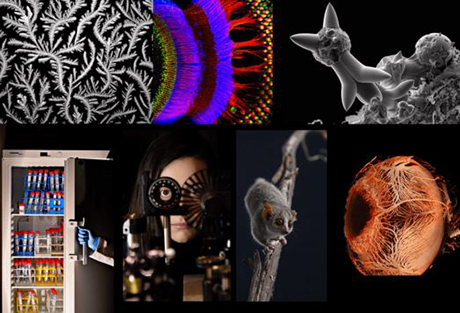
SNSF Scientific Image Competition
The SNSF Scientific Image Competition encourages researchers working in Switzerland to present their works to the public and the media. Photographs, images and videos will be rated in terms of their aesthetic quality and their ability to inspire and amaze, to convey or illustrate knowledge, to tell a human story or to let us discover a new universe. Submission deadline January 31, 2022. More information here.
PhD Defense
Federico Saltarin student ot the PhD program Cutting Edge Microscopy (CEM), will give his PhD defense on December 10, 2021 at 10.30 online via Zoom.
Title: “Investigations on the extravasation of melanoma cells across the blood brain barrier”.
Please request access to this online event via e-mail to Rahel Eyer.
Our project focuses on the investigation of the extravasation of melanoma cells across the blood brain barrier in the context of brain metastasis development.
To this aim, we addressed the impact of compromised endothelial junctions at the BBB on brain metastatic melanoma cell extravasation and we developed methods and tools for the investigation of melanoma brain metastasis formation on multi-scale level.
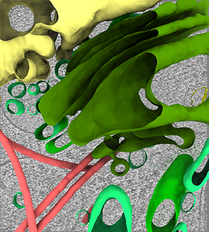
MIC Symposium 2021
Finally, after a missing year 2020 due to covid pandemic, the traditional MIC Symposium took place in UniS on November 19, 2021. The topic of this year was 3D electron microscopy. The scientific committee represented by Wanda Kukulski, Michael Stoffel, Benoît Zuber (University of Bern) and Dimitri Vanhecke (University of Fribourg), prepared an interesting program, which attracted more than 110 participants. More details see here.
LS2 Intersection Microscopy
The LS2 Intersection Microscopy together with the Swiss Society for Optics and Microscopy (SSOM) was founded in February 2019. During the past years, microscopy techniques as well as image analysis capabilities have developed at incredible pace. Today, microscopy based research is becoming increasingly important in all fields of life sciences. The LS2 microscopy intersection aims for a better and intensified knowledge transfer between the microscopy community and life scientists in Switzerland and thus intends to promote the faster introduction of new and cutting-edge microscopy techniques in Switzerland. Also, high-end microscopy techniques should become even better known as well as the core facilities providing them.
Apply for the LS2 membership here and select "Microscopy" in the online application form.
PhD Defense
Martin Liptay student ot the PhD program Cutting Edge Microscopy (CEM), will give his PhD defense on October 1, 2021 at 15:00 online via Zoom.
Title: “Replication fork remodeling and PARP inhibitor resistance in Brca1/2-mutated mouse mammary tumors”.
Please request access to this online event via e-mail to Rahel Eyer.
Poly (ADP-ribose) polymerase inhibitors (PARPi) are a class of targeted therapeutics that recently entered the clinic for the treatment of patients suffering from BRCA1/2-mutated breast and ovarian cancer. Unfortunately, similar to other anticancer therapeutics, their efficacy in tumor eradication is frequently limited by acquired drug resistance. By employing a variety of molecular biology and imaging techniques, we identified novel, non-canonical functions of DNA repair proteins H2AX and MDC1 in the metabolism of replication forks and in mediating PARPi response in BRCA1/2-deficient mammary tumors.
Zietschmann-Preuss-Award 2021
The 2021 Zietzschmann-Preuss Award presented by the World Association of Veterinary Anatomists goes to Christina Alexandra Kalenberg, Department of Clinical Veterinary Science, University of Bern, post-graduate student of MIC Board member Michael Stoffel. We congratulate her on this success!
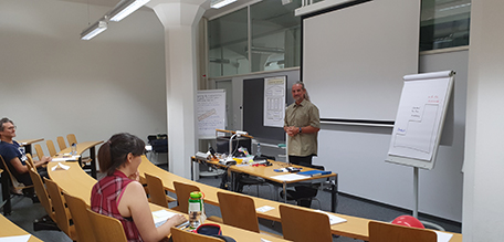
Communication training for students of the PhD program Cutting Edge Microscopy (CEM)
The communication training for CEM students took place on 6 and 8 September 2021 in Berne. During two interesting and varied days, the participants had the opportunity to work on impactful speaking techniques, exercices for voice warming and were able to improve their personal skills under instruction of Michael Berndonner, external public speaking and communication trainer.
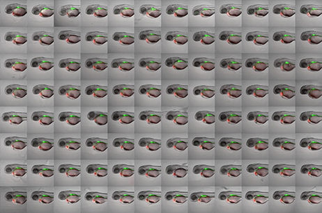
PhD Defense
Eleonora Lupi, student ot the PhD program Cutting Edge Microscopy (CEM), will give her PhD defense on September 29, 2021 at 15:30 online via Zoom.
Title: “Establishment of a zebrafish screening assay for the identification of compounds affecting proepicardial and epicardial formation”.
Please request access to this online event via e-mail to Rahel Eyer.
Description: the epicardium is the most external layer of the heart that has been demonstrated to have an important role during heart formation and regeneration. Despite the increasing interesting on this tissue, some mechanisms leading its formation remain still unknown. With this project we aimed to study the effect of epigenetic modulators on the proepicardial and epicardial formation in zebrafish model, to better understand the epigenetic role in this process. A drug screening was performed and zebrafish embryos were imaged using an Acquifer imaging machine. We were able to implement an automatic pipeline to quantify the number of the epicardial cells and determine the effect of the drugs on the epicardium.
PhD Defense
Alberto Mattei, student ot the PhD program Cutting Edge Microscopy (CEM), will give his PhD defense on September 1, 2021 at 10.00 am online via Zoom
Title: "Exploring Signaling Mechanisms regulating genetic and non-genetic Drug Resistance in Melanoma"
Please request access to this online event via e-mail to Rahel Eyer.
"Metastatic melanoma is a very aggressive type of cancer that in over 50% of cases carries a BRAFV600E mutation. The mutated BRAF drives aberrant MAPK-ERK pathway activation and tumor progression. Selective BRAFV600E inhibitors have been on the market since 2011, but drug resistance almost invariably develops and patient relapse in the clinic. We explored mechanisms that regulate genetic and non-genetic drug resistance in melanoma."
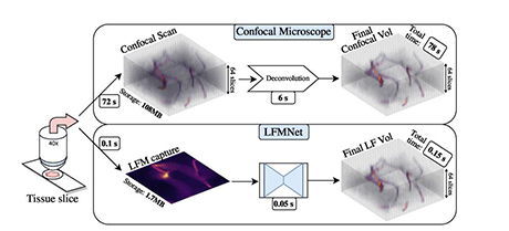
Congratulations to new publication!
Congratulations to Yury Belyaev, Federico Saltarin, Ruth Lyck and Paolo Favaro from University of Bern and their collaborators to their new publication on "Learning to Reconstruct Confocal Microscopy Stacks From Single Light Field Images" published in IEEE Transactions on Computational Imaging in July 2021, DOI.
Swiss Microscopy Core Facility Day 2021
Registration for the 2021 Swiss Microscopy Core Facility Day at the new Biozentrum in Basel is open.
Link to registration here.
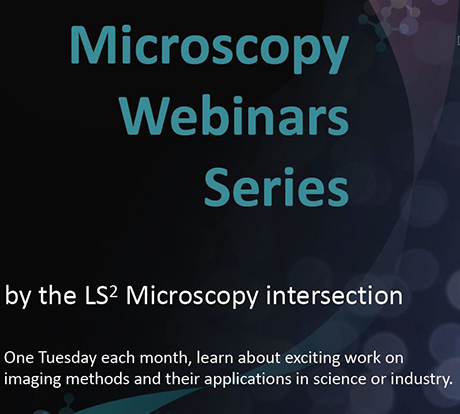
LS2 Webinar Series
The LS2 Microscopy Intersection is happy to organise the LS2 Microscopy Webinar Series. One Tuesday each month, a speaker will share their exciting work on imaging methods and their applications in science or industry.
The next LS2 Microscopy Webinar will take place on the 20th July 2021 at 17:00 (~30min plus Q&A time). Register here.
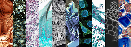
ZEISS Microscopy Image Contest 2021
We are thrilled to announce the ZEISS Microscopy Image Contest 2021. Do you take stunning microscopy images? Submit your best image now! The contest is open to all microscopy enthusiasts and professionals in any application field. Images can be taken with any ZEISS microscope (light, X-ray or electron microscope). Deadline for entry is July 31, 2021.
All participants will receive a copy of the ZEISS Microscopy Calendar 2022. On top of that, the three winners will receive the following prizes:
- 1st Prize: ZEISS Primostar 3
- 2nd Prize: ZEISS Victory Pocket 10x25
- 3rd Prize: ZEISS Terra 10x42
To submit your image and read all FAQs and rules, visit our contest web page
New position in microscopy at Wyss Center, Geneva
The position is conceived to focus on transversal activities among the different microscopes and image analysis systems with technical roles on maintenance, assistance and quality control of the microscopes and integration of image analysis pipelines. The person will also be involved in the run of Wyss Center driven research projects.
Deadline 23th of July, with possibility of extension.
Exciting publication of Professor Olivier Pertz
We congratulate MIC committee member Oliver Pertz and his team to their exciting new study “Collective ERK/Akt activity waves orchestrate epithelial homeostasis by driving apoptosis-induced survival” published in Developmental Cell in June 2021.
QUAREP-LiMi WG 10 Image Quality Talk from Alan C. Bovik
"On Picture Quality Prediction"
Alan Bovik is a faculty member in the Department of Electrical and Computer Engineering, the Wireless Networking and Communication Group (WNCG), and the Institute for Neuroscience. His research interests include digital television, digital photography, visual perception, social media, and image and video processing. To recieve the Zoom link to this talk, please contact Yury Belyaev.
15th EXCITE Zurich Summer School
Registration is now open for the 15th EXCITE Zurich Summer School on Biomedical Imaging that will take place in Zurich, Switzerland, from 6 - 17 September 2021. Application deadline: Monday, 3 May 2021 (Notification of acceptance: 1 June 2021).
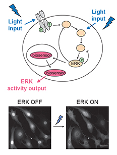
PhD Defense
Coralie Dessauges, student ot the PhD program Cutting Edge Microscopy (CEM), will give her PhD defense on May 3, 2021, at 1.00 pm. Title: An optogenetic actuator/biosensor circuit for large scale interrogation of MAPK signaling dynamics
Please request access to this online event via e-mail to Rahel Eyer.
In this work, we developed a set of optogenetic circuits to activate the MAPK/ERK pathway with light from different levels of the MAPK network and measure the corresponding ERK activity with a high temporal resolution. Together with automated imaging and image-analysis pipelines, our systems enable us to image and stimulate ERK signaling in hundreds of cells in a highly automated and dynamic fashion. Taking advantage of this throughput, we conducted an RNAi perturbation screen to investigate the role of 50 proteins of the MAPK network in ERK signaling regulation.
PhD Defense
Nora Ruef, student ot the PhD program Cutting Edge Microscopy (CEM), will give her PhD defense on March 26, 2021, at 4.00 pm. Please request access to this online event via e-mail to Rahel Eyer.
"In this thesis we investigated the unique migration mode of submandibular salivary gland tissue resident memory CD8+ T cells and how memory CD8+ T cells perform homeostatic surveillance of a complex organ with mixed epithelial – connective tissue composition."
PhD Defense
Julia Bruggisser, student of he PhD program Cutting Edge Microscopy (CEM), will give her PhD defense on March 25, 2021, at 3.00 pm. Please request access to this online event via e-mail to Rahel Eyer.
Title: Clostridium Perfringens β-Toxin: Receptor Identification and Oligomer Structure
Self-evaluation of the Microscopy Imaging Center (MIC)
Please complete the survey of the MIC here until March 16, 2021. This survey is designed to evaluate the performance of the MIC with respect to teaching and services. Your participation will help to implement improvements exactly there where you requested them most
New instrument at ARTORG
02.02.2021 - The newly acquired EVOS M7000 microscope from Thermo Fisher is a widefield fluorescence microscope, suitable for.... continue reading
White Paper of QUAREP-LiMi is online
29.01.2021 – Read the White Paper «QUAREP-LiMi: A community-driven initiative to establish guidelines for quality assessment and reproducibility for instruments and images in light microscopy» here.
Virtual Seminar Invitation - ImageXpress Pico
02.03.2020 - When size doesn't matter: meet ImageXpress Pico, the miniature but mighty microscope. Continue reading
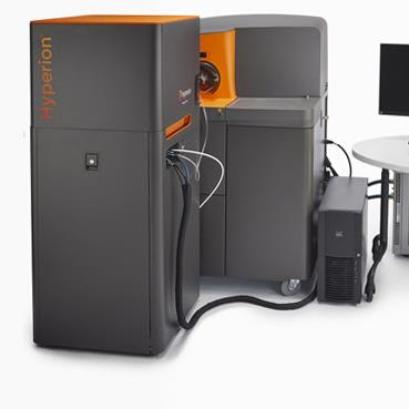
DBMR Hyperion Imaging System
02.11.20 – New MIC Instruments
Location: INO-F608A
Type: Imaging mass cytometer
Site: UniBE-Insel Read more

DBMR Helios Mass Cytometer
07.10.20 – New MIC Instruments
Location: INO-F608A
Type: Mass Cytometer
Site: UniBE-Insel Read more
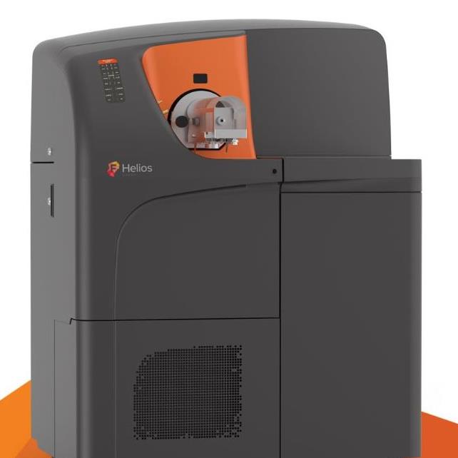
DBMR Hyperion Imaging System
05.10.20 – New MIC Instruments
Location: INO-F608A
Type: Imaging mass cytometer
Site: UniBE-Insel Read more
LHEP Keyence VHX-7000
05.10.20 – New MIC Instruments
Location: Gebäude der exakten Wissenschaften (ExWi)
Type: Widefield Microscope
Site: UniBE-ExWi Read more
IZB Nanolive 3D Cell Explorer
10.08.20 – New MIC Instruments
Location: Institute of Cell Biology, Baltzerstrasse 4, 3012 Bern
Type: Widefield Microscope
Site: UniBE-Muesmatt Read more
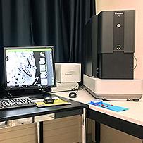
IRM ThermoFisher Phenom XL Scanning EM (Bob)
02.11.20 – New MIC Instruments
Location: Institute of Forensic Medicine, Bühlstrasse 20, 3012 Bern
Type: Electron Microscope
Site: UniBE-Muesmatt Read more